6XDG | pdb_00006xdg

Complex of SARS-CoV-2 receptor binding domain with the Fab fragments of two neutralizing antibodies


Experimental Data Snapshot

  • Method: ELECTRON MICROSCOPY
  • Resolution: 3.90 Å
  • Aggregation State: PARTICLE 
  • Reconstruction Method: SINGLE PARTICLE 

wwPDB Validation 3D Report Full Report

Validation slider image for 6XDG

This is version 2.3 of the entry. See complete history

Literature

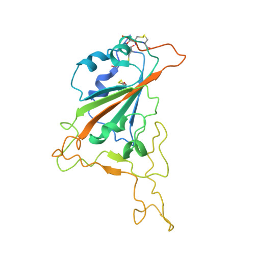
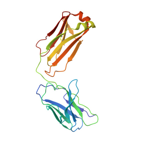
Studies in humanized mice and convalescent humans yield a SARS-CoV-2 antibody cocktail.

Hansen, J.Baum, A.Pascal, K.E.Russo, V.Giordano, S.Wloga, E.Fulton, B.O.Yan, Y.Koon, K.Patel, K.Chung, K.M.Hermann, A.Ullman, E.Cruz, J.Rafique, A.Huang, T.Fairhurst, J.Libertiny, C.Malbec, M.Lee, W.Y.Welsh, R.Farr, G.Pennington, S.Deshpande, D.Cheng, J.Watty, A.Bouffard, P.Babb, R.Levenkova, N.Chen, C.Zhang, B.Romero Hernandez, A.Saotome, K.Zhou, Y.Franklin, M.Sivapalasingam, S.Lye, D.C.Weston, S.Logue, J.Haupt, R.Frieman, M.Chen, G.Olson, W.Murphy, A.J.Stahl, N.Yancopoulos, G.D.Kyratsous, C.A.

(2020) Science 369: 1010-1014

  • DOI: https://doi.org/10.1126/science.abd0827
  • Primary Citation Related Structures: 
    6XDG

  • PubMed Abstract: 

    Neutralizing antibodies have become an important tool in treating infectious diseases. Recently, two separate approaches yielded successful antibody treatments for Ebola-one from genetically humanized mice and the other from a human survivor. Here, we describe parallel efforts using both humanized mice and convalescent patients to generate antibodies against the severe acute respiratory syndrome coronavirus 2 (SARS-CoV-2) spike protein, which yielded a large collection of fully human antibodies that were characterized for binding, neutralization, and three-dimensional structure. On the basis of these criteria, we selected pairs of highly potent individual antibodies that simultaneously bind the receptor binding domain of the spike protein, thereby providing ideal partners for a therapeutic antibody cocktail that aims to decrease the potential for virus escape mutants that might arise in response to selective pressure from a single-antibody treatment.


  • Organizational Affiliation
    • Regeneron Pharmaceuticals, Inc., Tarrytown, NY 10591, USA.

Macromolecule Content 

  • Total Structure Weight: 123.52 kDa 
  • Atom Count: 8,004 
  • Modeled Residue Count: 1,043 
  • Deposited Residue Count: 1,132 
  • Unique protein chains: 5

Macromolecules

Find similar proteins by:|  3D Structure
Entity ID: 1
MoleculeChains  Sequence LengthOrganismDetailsImage
Spike protein S1A [auth E]251Severe acute respiratory syndrome coronavirus 2Mutation(s): 0 
Gene Names: S2
UniProt
Find proteins for P0DTC2 (Severe acute respiratory syndrome coronavirus 2)
Explore P0DTC2 
Go to UniProtKB:  P0DTC2
Entity Groups
Sequence Clusters30% Identity50% Identity70% Identity90% Identity95% Identity100% Identity
UniProt GroupP0DTC2
Glycosylation
Glycosylation Sites: 1Go to GlyGen: P0DTC2-1
Sequence Annotations
Expand
Reference Sequence
Find similar proteins by:|  3D Structure
Entity ID: 2
MoleculeChains  Sequence LengthOrganismDetailsImage
REGN10933 antibody Fab fragment light chainB [auth D]214Homo sapiensMutation(s): 0 
Find similar proteins by:|  3D Structure
Entity ID: 3
MoleculeChains  Sequence LengthOrganismDetailsImage
REGN10933 antibody Fab fragment heavy chainC [auth B]226Homo sapiensMutation(s): 0 
Find similar proteins by:|  3D Structure
Entity ID: 4
MoleculeChains  Sequence LengthOrganismDetailsImage
REGN10987 antibody Fab fragment heavy chainD [auth C]225Homo sapiensMutation(s): 0 
Find similar proteins by:|  3D Structure
Entity ID: 5
MoleculeChains  Sequence LengthOrganismDetailsImage
REGN10987 antibody Fab fragment light chainE [auth A]216Homo sapiensMutation(s): 0 

Oligosaccharides

Help  
Entity ID: 6
MoleculeChains Length2D Diagram GlycosylationD Interactions
alpha-D-mannopyranose-(1-6)-beta-D-mannopyranose-(1-4)-2-acetamido-2-deoxy-beta-D-glucopyranose-(1-4)-2-acetamido-2-deoxy-beta-D-glucopyranose
F
4N-Glycosylation
Glycosylation Resources
GlyTouCan: G22573RC
GlyCosmos: G22573RC
GlyGen: G22573RC

Experimental Data & Validation

Experimental Data

  • Method: ELECTRON MICROSCOPY
  • Resolution: 3.90 Å
  • Aggregation State: PARTICLE 
  • Reconstruction Method: SINGLE PARTICLE 
EM Software:
TaskSoftware PackageVersion
RECONSTRUCTIONcryoSPARC
MODEL REFINEMENTPHENIX

Structure Validation

View Full Validation Report


Entry History 

& Funding Information

Deposition Data


Funding OrganizationLocationGrant Number
Biomedical Advanced Research and Development Authority (BARDA)HHSO100201700020C

Revision History  (Full details and data files)

  • Version 1.0: 2020-06-24
    Type: Initial release
  • Version 1.1: 2020-07-08
    Changes: Author supporting evidence
  • Version 1.2: 2020-07-22
    Changes: Source and taxonomy
  • Version 2.0: 2020-07-29
    Type: Remediation
    Reason: Carbohydrate remediation
    Changes: Advisory, Atomic model, Data collection, Derived calculations, Structure summary
  • Version 2.1: 2020-09-02
    Changes: Database references, Structure summary
  • Version 2.2: 2021-01-27
    Changes: Structure summary
  • Version 2.3: 2024-11-13
    Changes: Data collection, Database references, Structure summary