6WVF | pdb_00006wvf

E.coli DsbB C104S with ubiquinone

  • Classification: MEMBRANE PROTEIN
  • Organism(s): Aequorea victoria, Escherichia coli
  • Expression System: Escherichia coli
  • Mutation(s): Yes 
  • Membrane Protein: Yes  OPMPDBTMMemProtMDmpstruc

  • Deposited: 2020-05-05 Released: 2021-01-13 
  • Deposition Author(s): Liu, S., Li, W.
  • Funding Organization(s): National Institutes of Health/National Heart, Lung, and Blood Institute (NIH/NHLBI), Other private, National Institutes of Health/National Eye Institute (NIH/NEI), National Institutes of Health/National Institute of General Medical Sciences (NIH/NIGMS)

Experimental Data Snapshot

  • Method: X-RAY DIFFRACTION
  • Resolution: 2.90 Å
  • R-Value Free: 
    0.286 (Depositor), 0.303 (DCC) 
  • R-Value Work: 
    0.259 (Depositor), 0.265 (DCC) 
  • R-Value Observed: 
    0.260 (Depositor) 

Starting Model: experimental
View more details

wwPDB Validation 3D Report Full Report

Validation slider image for 6WVF

Ligand Structure Quality Assessment 


This is version 2.0 of the entry. See complete history

Literature

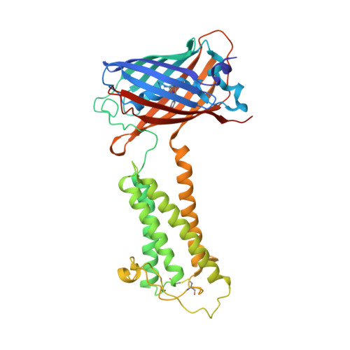
Termini restraining of small membrane proteins enables structure determination at near-atomic resolution.

Liu, S.Li, S.Yang, Y.Li, W.

(2020) Sci Adv 6

  • DOI: https://doi.org/10.1126/sciadv.abe3717
  • Primary Citation Related Structures: 
    6WVD, 6WVE, 6WVF

  • PubMed Abstract: 

    Small membrane proteins are difficult targets for structural characterization. Here, we stabilize their folding by restraining their amino and carboxyl termini with associable protein entities, exemplified by the two halves of a superfolder GFP. The termini-restrained proteins are functional and show improved stability during overexpression and purification. The reassembled GFP provides a versatile scaffold for membrane protein crystallization, enables diffraction to atomic resolution, and facilitates crystal identification, phase determination, and density modification. This strategy gives rise to 14 new structures of five vertebrate proteins from distinct functional families, bringing a substantial expansion to the structural database of small membrane proteins. Moreover, a high-resolution structure of bacterial DsbB reveals that this thiol oxidoreductase is activated through a catalytic triad, similar to cysteine proteases. Overall, termini restraining proves exceptionally effective for stabilization and structure determination of small membrane proteins.


  • Organizational Affiliation
    • Department of Biochemistry and Molecular Biophysics, Washington University School of Medicine, St. Louis, MO 63110, USA.

Macromolecule Content 

  • Total Structure Weight: 45.66 kDa 
  • Atom Count: 3,107 
  • Modeled Residue Count: 387 
  • Deposited Residue Count: 399 
  • Unique protein chains: 1

Macromolecules

Find similar proteins by:|  3D Structure
Entity ID: 1
MoleculeChains  Sequence LengthOrganismDetailsImage
Green fluorescent protein,Disulfide bond formation protein B,Green fluorescent protein399Aequorea victoriaEscherichia coliMutation(s): 4 
Gene Names: GFPdsbBroxBycgAb1185JW5182
Membrane Entity: Yes 
UniProt
Find proteins for P0A6M2 (Escherichia coli (strain K12))
Explore P0A6M2 
Go to UniProtKB:  P0A6M2
Find proteins for P42212 (Aequorea victoria)
Explore P42212 
Go to UniProtKB:  P42212
Entity Groups
Sequence Clusters30% Identity50% Identity70% Identity90% Identity95% Identity100% Identity
UniProt GroupsP42212P0A6M2
Sequence Annotations
Expand
Reference Sequence

Small Molecules

Ligands 1 Unique
IDChains Name / Formula / InChI Key2D Diagram3D Interactions
UQ1
(Subject of Investigation/LOI)

Query on UQ1


Download:Ideal Coordinates CCD File
B [auth A]UBIQUINONE-1
C14 H18 O4
SOECUQMRSRVZQQ-UHFFFAOYSA-N
Modified Residues  1 Unique
IDChains TypeFormula2D DiagramParent
CRO
Query on CRO
A
L-PEPTIDE LINKINGC15 H17 N3 O5THR, TYR, GLY

Experimental Data & Validation

Experimental Data

  • Method: X-RAY DIFFRACTION
  • Resolution: 2.90 Å
  • R-Value Free:  0.286 (Depositor), 0.303 (DCC) 
  • R-Value Work:  0.259 (Depositor), 0.265 (DCC) 
  • R-Value Observed: 0.260 (Depositor) 
Space Group: P 32 2 1
Unit Cell:
Length ( Å )Angle ( ˚ )
a = 53.381α = 90
b = 53.381β = 90
c = 280.74γ = 120
Software Package:
Software NamePurpose
Aimlessdata scaling
PHENIXrefinement
PDB_EXTRACTdata extraction
HKL-2000data reduction
PHASERphasing

Structure Validation

View Full Validation Report


Ligand Structure Quality Assessment 


Entry History 

& Funding Information

Deposition Data

  • Released Date: 2021-01-13 
  • Deposition Author(s): Liu, S., Li, W.

Funding OrganizationLocationGrant Number
National Institutes of Health/National Heart, Lung, and Blood Institute (NIH/NHLBI)United StatesR01 HL121718
Other privateUnited StatesForefront of Science Award
Other privateUnited StatesMCII 2020-854
National Institutes of Health/National Eye Institute (NIH/NEI)United StatesR21 EY028705
National Institutes of Health/National Institute of General Medical Sciences (NIH/NIGMS)United StatesR01 GM131008
National Institutes of Health/National Institute of General Medical Sciences (NIH/NIGMS)United StatesP30 GM124165

Revision History  (Full details and data files)

  • Version 1.0: 2021-01-13
    Type: Initial release
  • Version 1.1: 2023-10-18
    Changes: Data collection, Database references, Refinement description
  • Version 1.2: 2023-11-15
    Changes: Data collection
  • Version 1.3: 2024-11-13
    Changes: Structure summary
  • Version 2.0: 2026-03-18
    Changes: Polymer sequence