6WRF

ClpX-ClpP complex bound to GFP-ssrA, recognition complex


Experimental Data Snapshot

  • Method: ELECTRON MICROSCOPY
  • Resolution: 3.14 Å
  • Aggregation State: PARTICLE 
  • Reconstruction Method: SINGLE PARTICLE 

wwPDB Validation   3D Report Full Report


This is version 1.1 of the entry. See complete history


Literature

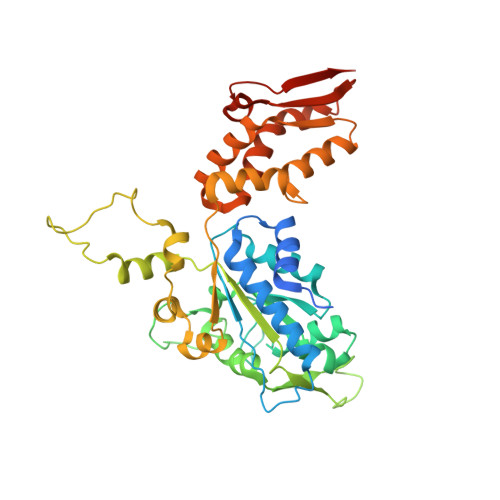
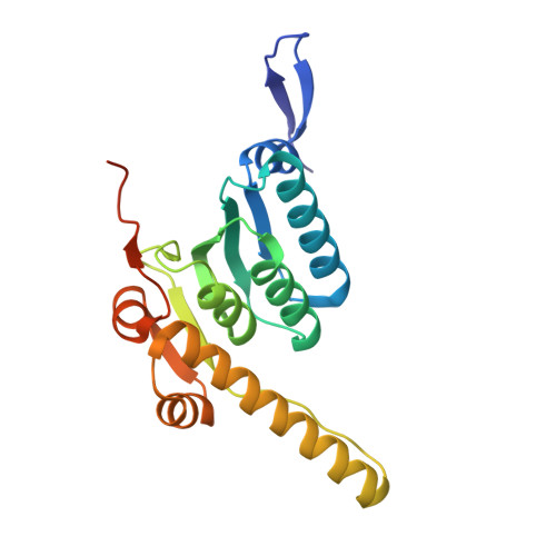
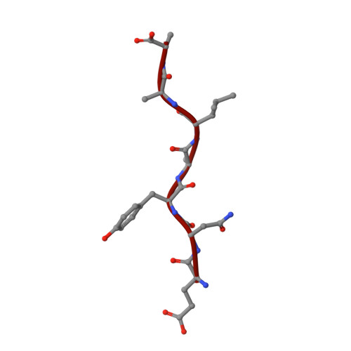
Structural basis of ClpXP recognition and unfolding of ssrA-tagged substrates.

Fei, X.Bell, T.A.Barkow, S.R.Baker, T.A.Sauer, R.T.

(2020) Elife 9

  • DOI: https://doi.org/10.7554/eLife.61496
  • Primary Citation of Related Structures:  
    6WR2, 6WRF, 6WSG

  • PubMed Abstract: 

    When ribosomes fail to complete normal translation, all cells have mechanisms to ensure degradation of the resulting partial proteins to safeguard proteome integrity. In Escherichia coli and other eubacteria, the tmRNA system rescues stalled ribosomes and adds an ssrA tag or degron to the C-terminus of the incomplete protein, which directs degradation by the AAA+ ClpXP protease. Here, we present cryo-EM structures of ClpXP bound to the ssrA degron. C-terminal residues of the ssrA degron initially bind in the top of an otherwise closed ClpX axial channel and subsequently move deeper into an open channel. For short-degron protein substrates, we show that unfolding can occur directly from the initial closed-channel complex. For longer degron substrates, our studies illuminate how ClpXP transitions from specific recognition into a nonspecific unfolding and translocation machine. Many AAA+ proteases and protein-remodeling motors are likely to employ similar multistep recognition and engagement strategies.


  • Organizational Affiliation

    Departments of Biology, Massachusetts Institute of Technology, Cambridge, United States.


Macromolecules
Find similar proteins by:  (by identity cutoff)  |  3D Structure
Entity ID: 1
MoleculeChains Sequence LengthOrganismDetailsImage
ATP-dependent Clp protease ATP-binding subunit ClpX388Escherichia coli K-12Mutation(s): 0 
Gene Names: clpXlopCb0438JW0428
UniProt
Find proteins for P0A6H1 (Escherichia coli (strain K12))
Explore P0A6H1 
Go to UniProtKB:  P0A6H1
Entity Groups  
Sequence Clusters30% Identity50% Identity70% Identity90% Identity95% Identity100% Identity
UniProt GroupP0A6H1
Sequence Annotations
Expand
  • Reference Sequence
Find similar proteins by:  (by identity cutoff)  |  3D Structure
Entity ID: 2
MoleculeChains Sequence LengthOrganismDetailsImage
ATP-dependent Clp protease proteolytic subunit207Escherichia coli K-12Mutation(s): 0 
Gene Names: clpPlopPb0437JW0427
EC: 3.4.21.92
UniProt
Find proteins for P0A6G7 (Escherichia coli (strain K12))
Explore P0A6G7 
Go to UniProtKB:  P0A6G7
Entity Groups  
Sequence Clusters30% Identity50% Identity70% Identity90% Identity95% Identity100% Identity
UniProt GroupP0A6G7
Sequence Annotations
Expand
  • Reference Sequence
Find similar proteins by:  (by identity cutoff)  |  3D Structure
Entity ID: 3
MoleculeChains Sequence LengthOrganismDetailsImage
Green fluorescent proteinM [auth S]274Aequorea victoriaMutation(s): 0 
Gene Names: GFP
UniProt
Find proteins for P42212 (Aequorea victoria)
Explore P42212 
Go to UniProtKB:  P42212
Entity Groups  
Sequence Clusters30% Identity50% Identity70% Identity90% Identity95% Identity100% Identity
UniProt GroupP42212
Sequence Annotations
Expand
  • Reference Sequence
Small Molecules
Ligands 3 Unique
IDChains Name / Formula / InChI Key2D Diagram3D Interactions
AGS
Query on AGS

Download Ideal Coordinates CCD File 
O [auth B],
Q [auth C],
S [auth D],
U [auth E],
X [auth A]
PHOSPHOTHIOPHOSPHORIC ACID-ADENYLATE ESTER
C10 H16 N5 O12 P3 S
NLTUCYMLOPLUHL-KQYNXXCUSA-N
ADP
Query on ADP

Download Ideal Coordinates CCD File 
W [auth F]ADENOSINE-5'-DIPHOSPHATE
C10 H15 N5 O10 P2
XTWYTFMLZFPYCI-KQYNXXCUSA-N
MG
Query on MG

Download Ideal Coordinates CCD File 
P [auth B],
R [auth C],
T [auth D],
V [auth E],
Y [auth A]
MAGNESIUM ION
Mg
JLVVSXFLKOJNIY-UHFFFAOYSA-N
Experimental Data & Validation

Experimental Data

  • Method: ELECTRON MICROSCOPY
  • Resolution: 3.14 Å
  • Aggregation State: PARTICLE 
  • Reconstruction Method: SINGLE PARTICLE 
EM Software:
TaskSoftware PackageVersion
MODEL REFINEMENTPHENIX1.14
RECONSTRUCTIONRELION3.0.8
RECONSTRUCTIONPHENIX1.14

Structure Validation

View Full Validation Report


Entry History & Funding Information

Deposition Data


Funding OrganizationLocationGrant Number
National Institutes of Health/National Institute of General Medical Sciences (NIH/NIGMS)United StatesGM-101988

Revision History  (Full details and data files)

  • Version 1.0: 2020-11-04
    Type: Initial release
  • Version 1.1: 2024-03-06
    Changes: Data collection, Database references, Refinement description