6WGH

Crystal structure of GDP-bound NRAS with ten residues long internal tandem duplication in the switch II region


Experimental Data Snapshot

  • Method: X-RAY DIFFRACTION
  • Resolution: 1.65 Å
  • R-Value Free: 0.209 
  • R-Value Work: 0.178 
  • R-Value Observed: 0.179 

wwPDB Validation   3D Report Full Report


Ligand Structure Quality Assessment 


This is version 1.3 of the entry. See complete history


Literature

RASinternal tandem duplication disrupts GTPase-activating protein (GAP) binding to activate oncogenic signaling.

Nelson, A.C.Turbyville, T.J.Dharmaiah, S.Rigby, M.Yang, R.Wang, T.Y.Columbus, J.Stephens, R.Taylor, T.Sciacca, D.Onsongo, G.Sarver, A.Subramanian, S.Nissley, D.V.Simanshu, D.K.Lou, E.

(2020) J Biol Chem 295: 9335-9348

  • DOI: https://doi.org/10.1074/jbc.RA119.011080
  • Primary Citation of Related Structures:  
    6PQ3, 6WGH

  • PubMed Abstract: 

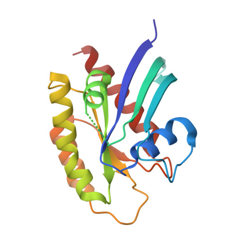
    The oncogene RAS is one of the most widely studied proteins in cancer biology, and mutant active RAS is a driver in many types of solid tumors and hematological malignancies. Yet the biological effects of different RAS mutations and the tissue-specific clinical implications are complex and nuanced. Here, we identified an internal tandem duplication (ITD) in the switch II domain of NRAS from a patient with extremely aggressive colorectal carcinoma. Results of whole-exome DNA sequencing of primary and metastatic tumors indicated that this mutation was present in all analyzed metastases and excluded the presence of any other clear oncogenic driver mutations. Biochemical analysis revealed increased interaction of the RAS ITD with Raf proto-oncogene Ser/Thr kinase (RAF), leading to increased phosphorylation of downstream MAPK/ERK kinase (MEK)/extracellular signal-regulated kinase (ERK). The ITD prevented interaction with neurofibromin 1 (NF1)-GTPase-activating protein (GAP), providing a mechanism for sustained activity of the RAS ITD protein. We present the first crystal structures of NRAS and KRAS ITD at 1.65-1.75 Å resolution, respectively, providing insight into the physical interactions of this class of RAS variants with its regulatory and effector proteins. Our in-depth bedside-to-bench analysis uncovers the molecular mechanism underlying a case of highly aggressive colorectal cancer and illustrates the importance of robust biochemical and biophysical approaches in the implementation of individualized medicine.


  • Organizational Affiliation

    Department of Laboratory Medicine & Pathology, University of Minnesota, Minneapolis, Minnesota, USA nels2055@umn.edu dhirendra.simanshu@nih.gov emil-lou@umn.edu.


Macromolecules
Find similar proteins by:  (by identity cutoff)  |  3D Structure
Entity ID: 1
MoleculeChains Sequence LengthOrganismDetailsImage
GTPase NRas
A, B
181Homo sapiensMutation(s): 0 
Gene Names: NRASHRAS1
UniProt & NIH Common Fund Data Resources
Find proteins for P01111 (Homo sapiens)
Explore P01111 
Go to UniProtKB:  P01111
PHAROS:  P01111
GTEx:  ENSG00000213281 
Entity Groups  
Sequence Clusters30% Identity50% Identity70% Identity90% Identity95% Identity100% Identity
UniProt GroupP01111
Sequence Annotations
Expand
  • Reference Sequence
Small Molecules
Ligands 4 Unique
IDChains Name / Formula / InChI Key2D Diagram3D Interactions
GDP (Subject of Investigation/LOI)
Query on GDP

Download Ideal Coordinates CCD File 
D [auth A],
I [auth B]
GUANOSINE-5'-DIPHOSPHATE
C10 H15 N5 O11 P2
QGWNDRXFNXRZMB-UUOKFMHZSA-N
FLC
Query on FLC

Download Ideal Coordinates CCD File 
E [auth A]CITRATE ANION
C6 H5 O7
KRKNYBCHXYNGOX-UHFFFAOYSA-K
GOL
Query on GOL

Download Ideal Coordinates CCD File 
F [auth A]GLYCEROL
C3 H8 O3
PEDCQBHIVMGVHV-UHFFFAOYSA-N
MG (Subject of Investigation/LOI)
Query on MG

Download Ideal Coordinates CCD File 
C [auth A],
G [auth A],
H [auth B]
MAGNESIUM ION
Mg
JLVVSXFLKOJNIY-UHFFFAOYSA-N
Experimental Data & Validation

Experimental Data

  • Method: X-RAY DIFFRACTION
  • Resolution: 1.65 Å
  • R-Value Free: 0.209 
  • R-Value Work: 0.178 
  • R-Value Observed: 0.179 
  • Space Group: P 1 21 1
Unit Cell:
Length ( Å )Angle ( ˚ )
a = 40.46α = 90
b = 45.96β = 99.77
c = 89.08γ = 90
Software Package:
Software NamePurpose
PHENIXrefinement
XDSdata reduction
XSCALEdata scaling
PHASERphasing
PDB_EXTRACTdata extraction

Structure Validation

View Full Validation Report


Ligand Structure Quality Assessment 


Entry History & Funding Information

Deposition Data


Funding OrganizationLocationGrant Number
National Institutes of Health/National Cancer Institute (NIH/NCI)United StatesHHSN261200800001E

Revision History  (Full details and data files)

  • Version 1.0: 2020-05-20
    Type: Initial release
  • Version 1.1: 2020-05-27
    Changes: Database references
  • Version 1.2: 2020-07-22
    Changes: Database references
  • Version 1.3: 2023-10-18
    Changes: Data collection, Database references, Derived calculations, Refinement description