Structural and spectroscopic characterization of photoactive yellow protein and photoswitchable fluorescent protein constructs containing heavy atoms.
Romei, M.G., Lin, C.Y., Boxer, S.G.(2020) J Photochem Photobiol A Chem 401: 112738
- PubMed: 32753830 Search on PubMedSearch on PubMed Central
- DOI: https://doi.org/10.1016/j.jphotochem.2020.112738
- Primary Citation Related Structures:
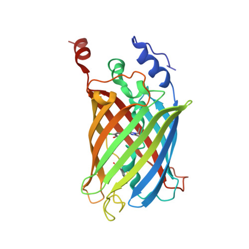
6UMY, 6UMZ, 6UN0, 6UN2, 6UN4 - PubMed Abstract:
Photo-induced structural rearrangements of chromophore-containing proteins are essential for various light-dependent signaling pathways and optogenetic applications. Ultrafast structural and spectroscopic methods have offered insights into these structural rearrangements across many timescales. However, questions still remain about exact mechanistic details, especially regarding photoisomerization of the chromophore within these proteins femtoseconds to picoseconds after photoexcitation. Instrumentation advancements for time-resolved crystallography and ultrafast electron diffraction provide a promising opportunity to study these reactions, but achieving enough signal-to-noise is a constant challenge. Here we present four new photoactive yellow protein constructs and one new fluorescent protein construct that contain heavy atoms either within or around the chromophore and can be expressed with high yields. Structural characterization of these constructs, most at atomic resolution, show minimal perturbation caused by the heavy atoms compared to wild-type structures. Spectroscopic studies report the effects of the heavy atom identity and location on the chromophore's photophysical properties. None of the substitutions prevent photoisomerization, although certain rates within the photocycle may be affected. Overall, these new proteins containing heavy atoms are ideal samples for state-of-theart time-resolved crystallography and electron diffraction experiments to elucidate crucial mechanistic information of photoisomerization.
- Department of Chemistry, Stanford University, Stanford, CA 94305, USA.
Organizational Affiliation: