A bifunctional O-antigen polymerase structure reveals a new glycosyltransferase family.
Clarke, B.R., Ovchinnikova, O.G., Sweeney, R.P., Kamski-Hennekam, E.R., Gitalis, R., Mallette, E., Kelly, S.D., Lowary, T.L., Kimber, M.S., Whitfield, C.(2020) Nat Chem Biol 16: 450-457
- PubMed: 32152541 Search on PubMedSearch on PubMed Central
- DOI: https://doi.org/10.1038/s41589-020-0494-0
- Primary Citation Related Structures:
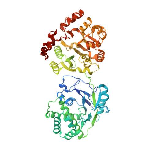
6U4B - PubMed Abstract:
Lipopolysaccharide O-antigen is an attractive candidate for immunotherapeutic strategies targeting antibiotic-resistant Klebsiella pneumoniae. Several K. pneumoniae O-serotypes are based on a shared O2a-antigen backbone repeating unit: (→ 3)-α-Galp-(1 → 3)-β-Galf-(1 →). O2a antigen is synthesized on undecaprenol diphosphate in a pathway involving the O2a polymerase, WbbM, before its export by an ATP-binding cassette transporter. This dual domain polymerase possesses a C-terminal galactopyranosyltransferase resembling known GT8 family enzymes, and an N-terminal DUF4422 domain identified here as a galactofuranosyltransferase defining a previously unrecognized family (GT111). Functional assignment of DUF4422 explains how galactofuranose is incorporated into various polysaccharides of importance in vaccine production and the food industry. In the 2.1-Å resolution structure, three WbbM protomers associate to form a flattened triangular prism connected to a central stalk that orients the active sites toward the membrane. The biochemical, structural and topological properties of WbbM offer broader insight into the mechanisms of assembly of bacterial cell-surface glycans.
- Department of Molecular and Cellular Biology, University of Guelph, Guelph, Ontario, Canada.
Organizational Affiliation: