6RQ7

Gadolinium MRI contrast compound binding in human plasma glycoprotein afamin - resurrection of highly anisotropic data


Experimental Data Snapshot

  • Method: X-RAY DIFFRACTION
  • Resolution: 2.69 Å
  • R-Value Free: 0.335 
  • R-Value Work: 0.263 
  • R-Value Observed: 0.266 

wwPDB Validation   3D Report Full Report


This is version 2.0 of the entry. See complete history


Literature

Controlled dehydration, structural flexibility and gadolinium MRI contrast compound binding in the human plasma glycoprotein afamin.

Naschberger, A.Juyoux, P.von Velsen, J.Rupp, B.Bowler, M.W.

(2019) Acta Crystallogr D Struct Biol 75: 1071-1083

  • DOI: https://doi.org/10.1107/S2059798319013500
  • Primary Citation of Related Structures:  
    6FAK, 6RQ7

  • PubMed Abstract: 

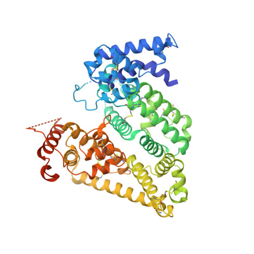
    Afamin, which is a human blood plasma glycoprotein, a putative multifunctional transporter of hydrophobic molecules and a marker for metabolic syndrome, poses multiple challenges for crystallographic structure determination, both practically and in analysis of the models. Several hundred crystals were analysed, and an unusual variability in cell volume and difficulty in solving the structure despite an ∼34% sequence identity with nonglycosylated human serum albumin indicated that the molecule exhibits variable and context-sensitive packing, despite the simplified glycosylation in insect cell-expressed recombinant afamin. Controlled dehydration of the crystals was able to stabilize the orthorhombic crystal form, reducing the number of molecules in the asymmetric unit from the monoclinic form and changing the conformational state of the protein. An iterative strategy using fully automatic experiments available on MASSIF-1 was used to quickly determine the optimal protocol to achieve the phase transition, which should be readily applicable to many types of sample. The study also highlights the drawback of using a single crystallographic structure model for computational modelling purposes given that the conformational state of the binding sites and the electron density in the binding site, which is likely to result from PEGs, greatly varies between models. This also holds for the analysis of nonspecific low-affinity ligands, where often a variety of fragments with similar uncertainty can be modelled, inviting interpretative bias. As a promiscuous transporter, afamin also seems to bind gadoteridol, a magnetic resonance imaging contrast compound, in at least two sites. One pair of gadoteridol molecules is located near the human albumin Sudlow site, and a second gadoteridol molecule is located at an intermolecular site in proximity to domain IA. The data from the co-crystals support modern metrics of data quality in the context of the information that can be gleaned from data sets that would be abandoned on classical measures.


  • Organizational Affiliation

    Department of Genetic Epidemiology, Medical University Innsbruck, Schöpfstrasse 41, A-6020 Innsbruck, Austria.


Macromolecules
Find similar proteins by:  (by identity cutoff)  |  3D Structure
Entity ID: 1
MoleculeChains Sequence LengthOrganismDetailsImage
AfaminA [auth B]586Homo sapiensMutation(s): 0 
Gene Names: AFMALB2ALBA
UniProt & NIH Common Fund Data Resources
Find proteins for P43652 (Homo sapiens)
Explore P43652 
Go to UniProtKB:  P43652
PHAROS:  P43652
GTEx:  ENSG00000079557 
Entity Groups  
Sequence Clusters30% Identity50% Identity70% Identity90% Identity95% Identity100% Identity
UniProt GroupP43652
Sequence Annotations
Expand
  • Reference Sequence
Oligosaccharides

Help

Entity ID: 2
MoleculeChains Length2D Diagram Glycosylation3D Interactions
2-acetamido-2-deoxy-beta-D-glucopyranose-(1-4)-2-acetamido-2-deoxy-beta-D-glucopyranoseB [auth A]2N-Glycosylation
Glycosylation Resources
GlyTouCan:  G42666HT
GlyCosmos:  G42666HT
GlyGen:  G42666HT
Small Molecules
Ligands 3 Unique
IDChains Name / Formula / InChI Key2D Diagram3D Interactions
DO3
Query on DO3

Download Ideal Coordinates CCD File 
H [auth B],
I [auth B],
J [auth B]
10-((2R)-2-HYDROXYPROPYL)-1,4,7,10-TETRAAZACYCLODODECANE 1,4,7-TRIACETIC ACID
C17 H32 N4 O7
IQUHNCOJRJBMSU-CQSZACIVSA-N
NAG
Query on NAG

Download Ideal Coordinates CCD File 
F [auth B],
G [auth B]
2-acetamido-2-deoxy-beta-D-glucopyranose
C8 H15 N O6
OVRNDRQMDRJTHS-FMDGEEDCSA-N
GD (Subject of Investigation/LOI)
Query on GD

Download Ideal Coordinates CCD File 
C [auth B],
D [auth B],
E [auth B]
GADOLINIUM ATOM
Gd
UIWYJDYFSGRHKR-UHFFFAOYSA-N
Experimental Data & Validation

Experimental Data

  • Method: X-RAY DIFFRACTION
  • Resolution: 2.69 Å
  • R-Value Free: 0.335 
  • R-Value Work: 0.263 
  • R-Value Observed: 0.266 
  • Space Group: P 21 21 2
Unit Cell:
Length ( Å )Angle ( ˚ )
a = 103.352α = 90
b = 109.734β = 90
c = 48.381γ = 90
Software Package:
Software NamePurpose
REFMACrefinement
XDSdata reduction
pointlessdata scaling
Aimlessdata scaling
autoPROCdata collection
STARANISOdata scaling
PHASERphasing

Structure Validation

View Full Validation Report


Entry History & Funding Information

Deposition Data


Funding OrganizationLocationGrant Number
Austrian Science FundAustriaP28395-B26

Revision History  (Full details and data files)

  • Version 1.0: 2019-07-10
    Type: Initial release
  • Version 1.1: 2019-12-18
    Changes: Database references
  • Version 1.2: 2020-07-08
    Changes: Data collection
  • Version 2.0: 2020-07-29
    Type: Remediation
    Reason: Carbohydrate remediation
    Changes: Atomic model, Data collection, Derived calculations, Refinement description, Structure summary