6RED | pdb_00006red

Cryo-EM structure of Polytomella F-ATP synthase, Rotary substate 3A, focussed refinement of F1 head and rotor


Experimental Data Snapshot

  • Method: ELECTRON MICROSCOPY
  • Resolution: 3.00 Å
  • Aggregation State: PARTICLE 
  • Reconstruction Method: SINGLE PARTICLE 

wwPDB Validation 3D Report Full Report

Validation slider image for 6RED

This is version 1.3 of the entry. See complete history

Literature

Rotary substates of mitochondrial ATP synthase reveal the basis of flexible F 1 -F o coupling.

Murphy, B.J.Klusch, N.Langer, J.Mills, D.J.Yildiz, O.Kuhlbrandt, W.

(2019) Science 364

  • DOI: https://doi.org/10.1126/science.aaw9128
  • Primary Citation Related Structures: 
    6RD4, 6RD5, 6RD6, 6RD7, 6RD8, 6RD9, 6RDA, 6RDB, 6RDC, 6RDD, 6RDE, 6RDF, 6RDG, 6RDH, 6RDI, 6RDJ, 6RDK, 6RDL, 6RDM, 6RDN, 6RDO, 6RDP, 6RDQ, 6RDR, 6RDS, 6RDT, 6RDU, 6RDV, 6RDW, 6RDX, 6RDY, 6RDZ, 6RE0, 6RE1, 6RE2, 6RE3, 6RE4, 6RE5, 6RE6, 6RE7, 6RE8, 6RE9, 6REA, 6REB, 6REC, 6RED, 6REE, 6REF, 6REP, 6RER, ... Search all related entries

  • PubMed Abstract: 

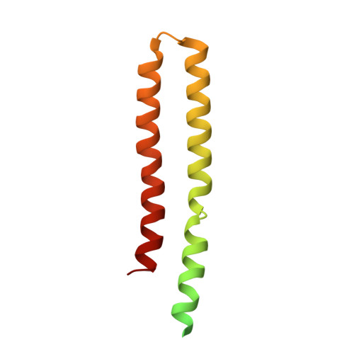
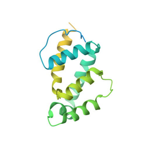
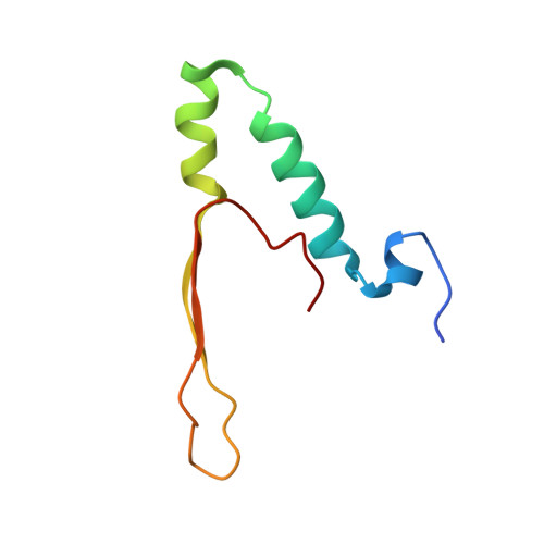
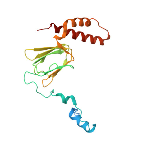
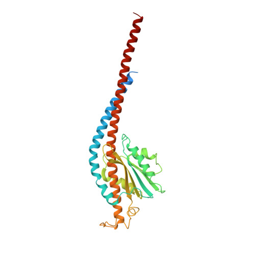
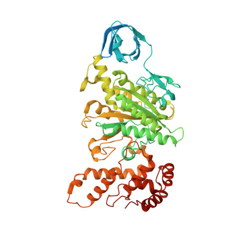
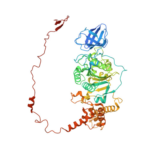
    F 1 F o -adenosine triphosphate (ATP) synthases make the energy of the proton-motive force available for energy-consuming processes in the cell. We determined the single-particle cryo-electron microscopy structure of active dimeric ATP synthase from mitochondria of Polytomella sp. at a resolution of 2.7 to 2.8 angstroms. Separation of 13 well-defined rotary substates by three-dimensional classification provides a detailed picture of the molecular motions that accompany c -ring rotation and result in ATP synthesis. Crucially, the F 1 head rotates along with the central stalk and c -ring rotor for the first ~30° of each 120° primary rotary step to facilitate flexible coupling of the stoichiometrically mismatched F 1 and F o subcomplexes. Flexibility is mediated primarily by the interdomain hinge of the conserved OSCP subunit. A conserved metal ion in the proton access channel may synchronize c -ring protonation with rotation.


  • Organizational Affiliation
    • Department of Structural Biology, Max Planck Institute of Biophysics, Frankfurt 60438, Germany.

Macromolecule Content 

  • Total Structure Weight: 586.51 kDa 
  • Atom Count: 33,899 
  • Modeled Residue Count: 4,503 
  • Deposited Residue Count: 5,497 
  • Unique protein chains: 7

Macromolecules

Find similar proteins by:|  3D Structure
Entity ID: 1
MoleculeChains  Sequence LengthOrganismDetailsImage
Mitochondrial ATP synthase subunit c
A, B, C, D, E
A, B, C, D, E, F, G, H, I, J
127Polytomella sp. Pringsheim 198.80Mutation(s): 0 
Membrane Entity: Yes 
UniProt
Find proteins for D7P7X5 (Polytomella sp. Pringsheim 198.80)
Explore D7P7X5 
Go to UniProtKB:  D7P7X5
Entity Groups
Sequence Clusters30% Identity50% Identity70% Identity90% Identity95% Identity100% Identity
UniProt GroupD7P7X5
Sequence Annotations
Expand
Reference Sequence
Find similar proteins by:|  3D Structure
Entity ID: 2
MoleculeChains  Sequence LengthOrganismDetailsImage
Mitochondrial ATP synthase subunit OSCPK [auth P]229Polytomella sp. Pringsheim 198.80Mutation(s): 0 
Membrane Entity: Yes 
UniProt
Find proteins for D8V7I1 (Polytomella sp. Pringsheim 198.80)
Explore D8V7I1 
Go to UniProtKB:  D8V7I1
Entity Groups
Sequence Clusters30% Identity50% Identity70% Identity90% Identity95% Identity100% Identity
UniProt GroupD8V7I1
Sequence Annotations
Expand
Reference Sequence
Find similar proteins by:|  3D Structure
Entity ID: 3
MoleculeChains  Sequence LengthOrganismDetailsImage
epsilon: Polytomella F-ATP synthase epsilon subunitL [auth Q]74Polytomella sp. Pringsheim 198.80Mutation(s): 0 
UniProt
Find proteins for A0A5H1ZR74 (Polytomella sp. Pringsheim 198.80)
Explore A0A5H1ZR74 
Go to UniProtKB:  A0A5H1ZR74
Entity Groups
Sequence Clusters30% Identity50% Identity70% Identity90% Identity95% Identity100% Identity
UniProt GroupA0A5H1ZR74
Sequence Annotations
Expand
Reference Sequence
Find similar proteins by:|  3D Structure
Entity ID: 4
MoleculeChains  Sequence LengthOrganismDetailsImage
Mitochondrial ATP synthase subunit deltaM [auth R]199Polytomella sp. Pringsheim 198.80Mutation(s): 0 
Membrane Entity: Yes 
UniProt
Find proteins for D7P7X6 (Polytomella sp. Pringsheim 198.80)
Explore D7P7X6 
Go to UniProtKB:  D7P7X6
Entity Groups
Sequence Clusters30% Identity50% Identity70% Identity90% Identity95% Identity100% Identity
UniProt GroupD7P7X6
Sequence Annotations
Expand
Reference Sequence
Find similar proteins by:|  3D Structure
Entity ID: 5
MoleculeChains  Sequence LengthOrganismDetailsImage
ATP synthase gamma chain, mitochondrialN [auth S]317Polytomella sp. Pringsheim 198.80Mutation(s): 0 
Membrane Entity: Yes 
UniProt
Find proteins for Q4LDE7 (Polytomella sp. Pringsheim 198.80)
Explore Q4LDE7 
Go to UniProtKB:  Q4LDE7
Entity Groups
Sequence Clusters30% Identity50% Identity70% Identity90% Identity95% Identity100% Identity
UniProt GroupQ4LDE7
Sequence Annotations
Expand
Reference Sequence
Find similar proteins by:|  3D Structure
Entity ID: 6
MoleculeChains  Sequence LengthOrganismDetailsImage
ATP synthase subunit alphaO [auth T],
P [auth U],
Q [auth V]
562Polytomella sp. Pringsheim 198.80Mutation(s): 0 
Membrane Entity: Yes 
UniProt
Find proteins for A0ZW40 (Polytomella sp. Pringsheim 198.80)
Explore A0ZW40 
Go to UniProtKB:  A0ZW40
Entity Groups
Sequence Clusters30% Identity50% Identity70% Identity90% Identity95% Identity100% Identity
UniProt GroupA0ZW40
Sequence Annotations
Expand
Reference Sequence
Find similar proteins by:|  3D Structure
Entity ID: 7
MoleculeChains  Sequence LengthOrganismDetailsImage
ATP synthase subunit betaR [auth X],
S [auth Y],
T [auth Z]
574Polytomella sp. Pringsheim 198.80Mutation(s): 0 
EC: 7.1.2.2
Membrane Entity: Yes 
UniProt
Find proteins for A0ZW41 (Polytomella sp. Pringsheim 198.80)
Explore A0ZW41 
Go to UniProtKB:  A0ZW41
Entity Groups
Sequence Clusters30% Identity50% Identity70% Identity90% Identity95% Identity100% Identity
UniProt GroupA0ZW41
Sequence Annotations
Expand
Reference Sequence

Small Molecules

Ligands 3 Unique
IDChains Name / Formula / InChI Key2D Diagram3D Interactions
ATP

Query on ATP


Download:Ideal Coordinates CCD File
U [auth T],
W [auth U],
Y [auth V]
ADENOSINE-5'-TRIPHOSPHATE
C10 H16 N5 O13 P3
ZKHQWZAMYRWXGA-KQYNXXCUSA-N
ADP

Query on ADP


Download:Ideal Coordinates CCD File
AA [auth X],
CA [auth Z]
ADENOSINE-5'-DIPHOSPHATE
C10 H15 N5 O10 P2
XTWYTFMLZFPYCI-KQYNXXCUSA-N
MG

Query on MG


Download:Ideal Coordinates CCD File
BA [auth X],
DA [auth Z],
V [auth T],
X [auth U],
Z [auth V]
MAGNESIUM ION
Mg
JLVVSXFLKOJNIY-UHFFFAOYSA-N

Experimental Data & Validation

Experimental Data

  • Method: ELECTRON MICROSCOPY
  • Resolution: 3.00 Å
  • Aggregation State: PARTICLE 
  • Reconstruction Method: SINGLE PARTICLE 
EM Software:
TaskSoftware PackageVersion
MODEL REFINEMENTPHENIX1.14
RECONSTRUCTIONRELION3

Structure Validation

View Full Validation Report


Entry History 

& Funding Information

Deposition Data


Funding OrganizationLocationGrant Number
Max Planck SocietyGermany--
European Molecular Biology OrganizationGermanyALTF 702 2016

Revision History  (Full details and data files)

  • Version 1.0: 2019-07-03
    Type: Initial release
  • Version 1.1: 2019-12-18
    Changes: Other
  • Version 1.2: 2020-09-30
    Changes: Database references, Derived calculations
  • Version 1.3: 2024-05-22
    Changes: Data collection, Database references