6R92

Cryo-EM structure of NCP-THF2(+1)-UV-DDB class B


Experimental Data Snapshot

  • Method: ELECTRON MICROSCOPY
  • Resolution: 4.80 Å
  • Aggregation State: PARTICLE 
  • Reconstruction Method: SINGLE PARTICLE 

Starting Models: experimental
View more details

wwPDB Validation   3D Report Full Report


This is version 1.4 of the entry. See complete history


Literature

DNA damage detection in nucleosomes involves DNA register shifting.

Matsumoto, S.Cavadini, S.Bunker, R.D.Grand, R.S.Potenza, A.Rabl, J.Yamamoto, J.Schenk, A.D.Schubeler, D.Iwai, S.Sugasawa, K.Kurumizaka, H.Thoma, N.H.

(2019) Nature 571: 79-84

  • DOI: https://doi.org/10.1038/s41586-019-1259-3
  • Primary Citation of Related Structures:  
    6R8Y, 6R8Z, 6R90, 6R91, 6R92, 6R93, 6R94

  • PubMed Abstract: 

    Access to DNA packaged in nucleosomes is critical for gene regulation, DNA replication and DNA repair. In humans, the UV-damaged DNA-binding protein (UV-DDB) complex detects UV-light-induced pyrimidine dimers throughout the genome; however, it remains unknown how these lesions are recognized in chromatin, in which nucleosomes restrict access to DNA. Here we report cryo-electron microscopy structures of UV-DDB bound to nucleosomes bearing a 6-4 pyrimidine-pyrimidone dimer or a DNA-damage mimic in various positions. We find that UV-DDB binds UV-damaged nucleosomes at lesions located in the solvent-facing minor groove without affecting the overall nucleosome architecture. In the case of buried lesions that face the histone core, UV-DDB changes the predominant translational register of the nucleosome and selectively binds the lesion in an accessible, exposed position. Our findings explain how UV-DDB detects occluded lesions in strongly positioned nucleosomes, and identify slide-assisted site exposure as a mechanism by which high-affinity DNA-binding proteins can access otherwise occluded sites in nucleosomal DNA.


  • Organizational Affiliation

    Friedrich Miescher Institute for Biomedical Research, Basel, Switzerland.


Macromolecules

Find similar proteins by:  (by identity cutoff)  |  3D Structure
Entity ID: 3
MoleculeChains Sequence LengthOrganismDetailsImage
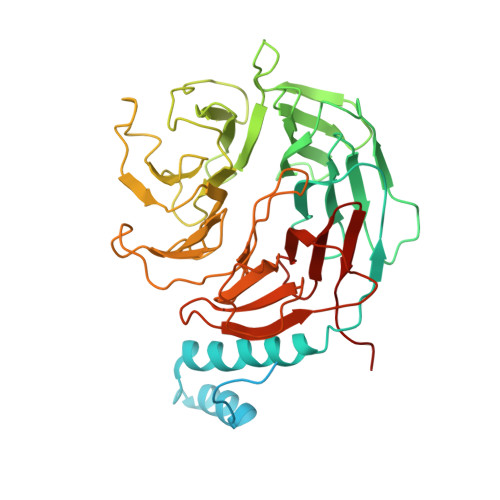
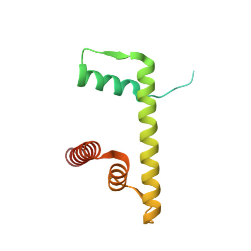
DNA damage-binding protein 2C [auth L]450Homo sapiensMutation(s): 0 
Gene Names: DDB2
UniProt & NIH Common Fund Data Resources
Find proteins for Q92466 (Homo sapiens)
Explore Q92466 
Go to UniProtKB:  Q92466
PHAROS:  Q92466
GTEx:  ENSG00000134574 
Entity Groups  
Sequence Clusters30% Identity50% Identity70% Identity90% Identity95% Identity100% Identity
UniProt GroupQ92466
Sequence Annotations
Expand
  • Reference Sequence
Find similar proteins by:  (by identity cutoff)  |  3D Structure
Entity ID: 4
MoleculeChains Sequence LengthOrganismDetailsImage
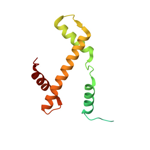
Histone H3.1D [auth A],
H [auth E]
139Homo sapiensMutation(s): 0 
Gene Names: 
UniProt & NIH Common Fund Data Resources
Find proteins for P68431 (Homo sapiens)
Explore P68431 
Go to UniProtKB:  P68431
PHAROS:  P68431
Entity Groups  
Sequence Clusters30% Identity50% Identity70% Identity90% Identity95% Identity100% Identity
UniProt GroupP68431
Sequence Annotations
Expand
  • Reference Sequence
Find similar proteins by:  (by identity cutoff)  |  3D Structure
Entity ID: 5
MoleculeChains Sequence LengthOrganismDetailsImage
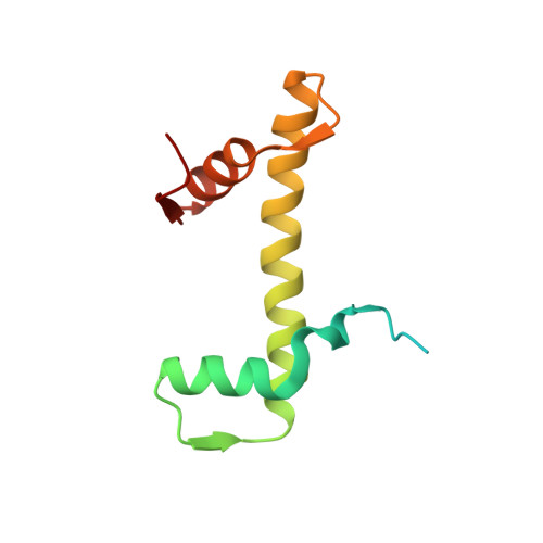
Histone H4E [auth B],
I [auth F]
106Homo sapiensMutation(s): 0 
Gene Names: 
UniProt & NIH Common Fund Data Resources
Find proteins for P62805 (Homo sapiens)
Explore P62805 
Go to UniProtKB:  P62805
PHAROS:  P62805
Entity Groups  
Sequence Clusters30% Identity50% Identity70% Identity90% Identity95% Identity100% Identity
UniProt GroupP62805
Sequence Annotations
Expand
  • Reference Sequence
Find similar proteins by:  (by identity cutoff)  |  3D Structure
Entity ID: 6
MoleculeChains Sequence LengthOrganismDetailsImage
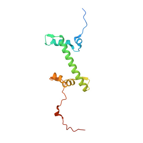
Histone H2A type 1-B/EF [auth C],
J [auth G]
133Homo sapiensMutation(s): 0 
Gene Names: HIST1H2ABH2AFMHIST1H2AEH2AFA
UniProt & NIH Common Fund Data Resources
Find proteins for P04908 (Homo sapiens)
Explore P04908 
Go to UniProtKB:  P04908
PHAROS:  P04908
Entity Groups  
Sequence Clusters30% Identity50% Identity70% Identity90% Identity95% Identity100% Identity
UniProt GroupP04908
Sequence Annotations
Expand
  • Reference Sequence
Find similar proteins by:  (by identity cutoff)  |  3D Structure
Entity ID: 7
MoleculeChains Sequence LengthOrganismDetailsImage
Histone H2B type 1-JG [auth D],
K [auth H]
129Homo sapiensMutation(s): 0 
Gene Names: HIST1H2BJH2BFR
UniProt & NIH Common Fund Data Resources
Find proteins for P06899 (Homo sapiens)
Explore P06899 
Go to UniProtKB:  P06899
PHAROS:  P06899
GTEx:  ENSG00000124635 
Entity Groups  
Sequence Clusters30% Identity50% Identity70% Identity90% Identity95% Identity100% Identity
UniProt GroupP06899
Sequence Annotations
Expand
  • Reference Sequence
Find similar proteins by:  (by identity cutoff)  |  3D Structure
Entity ID: 8
MoleculeChains Sequence LengthOrganismDetailsImage
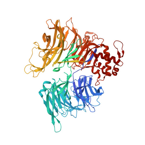
DNA damage-binding protein 1,DNA damage-binding protein 1L [auth K]1,163Homo sapiensMutation(s): 0 
Gene Names: DDB1XAP1
UniProt & NIH Common Fund Data Resources
Find proteins for Q16531 (Homo sapiens)
Explore Q16531 
Go to UniProtKB:  Q16531
PHAROS:  Q16531
GTEx:  ENSG00000167986 
Entity Groups  
Sequence Clusters30% Identity50% Identity70% Identity90% Identity95% Identity100% Identity
UniProt GroupQ16531
Sequence Annotations
Expand
  • Reference Sequence
Find similar nucleic acids by:  (by identity cutoff)  |  3D Structure
Entity ID: 1
MoleculeChains LengthOrganismImage
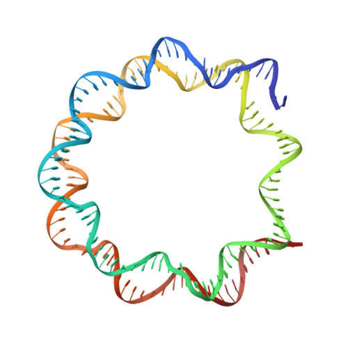
Human alpha-satellite DNA (145-MER)A [auth I]145Homo sapiens
Sequence Annotations
Expand
  • Reference Sequence
Find similar nucleic acids by:  (by identity cutoff)  |  3D Structure
Entity ID: 2
MoleculeChains LengthOrganismImage
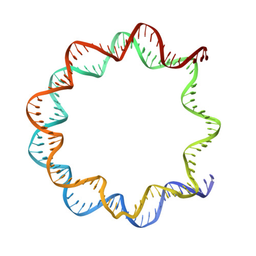
Human alpha-satellite DNA (145-MER) with abasic sites at positions 93-94B [auth J]145Homo sapiens
Sequence Annotations
Expand
  • Reference Sequence
Experimental Data & Validation

Experimental Data

  • Method: ELECTRON MICROSCOPY
  • Resolution: 4.80 Å
  • Aggregation State: PARTICLE 
  • Reconstruction Method: SINGLE PARTICLE 
EM Software:
TaskSoftware PackageVersion
RECONSTRUCTIONRELION2
MODEL REFINEMENTREFMAC5.8.0238
MODEL REFINEMENTPHENIXdev-3318

Structure Validation

View Full Validation Report


Entry History & Funding Information

Deposition Data


Funding OrganizationLocationGrant Number
Swiss National Science FoundationSwitzerlandCRSII3_160734/1
European Union666068
European Commission667951
European Commission705354
Japan Society for the Promotion of ScienceJapanJP18H05534
Japan Society for the Promotion of ScienceJapanJP16H06307
Japan Agency for Medical Research and Development (AMED)JapanJP18am0101076

Revision History  (Full details and data files)

  • Version 1.0: 2019-06-12
    Type: Initial release
  • Version 1.1: 2019-07-17
    Changes: Author supporting evidence, Data collection, Database references
  • Version 1.2: 2019-11-06
    Changes: Data collection, Refinement description
  • Version 1.3: 2019-12-18
    Changes: Other
  • Version 1.4: 2024-05-22
    Changes: Data collection, Database references, Refinement description