6QGI

Crystal structure of VP5 from Haloarchaeal pleomorphic virus 2


Experimental Data Snapshot

  • Method: X-RAY DIFFRACTION
  • Resolution: 2.46 Å
  • R-Value Free: 0.241 
  • R-Value Work: 0.224 
  • R-Value Observed: 0.225 

wwPDB Validation   3D Report Full Report


Ligand Structure Quality Assessment 


This is version 1.1 of the entry. See complete history


Literature

The structure of a prokaryotic viral envelope protein expands the landscape of membrane fusion proteins.

El Omari, K.Li, S.Kotecha, A.Walter, T.S.Bignon, E.A.Harlos, K.Somerharju, P.De Haas, F.Clare, D.K.Molin, M.Hurtado, F.Li, M.Grimes, J.M.Bamford, D.H.Tischler, N.D.Huiskonen, J.T.Stuart, D.I.Roine, E.

(2019) Nat Commun 10: 846-846

  • DOI: https://doi.org/10.1038/s41467-019-08728-7
  • Primary Citation of Related Structures:  
    6J7V, 6QGI, 6QGL

  • PubMed Abstract: 

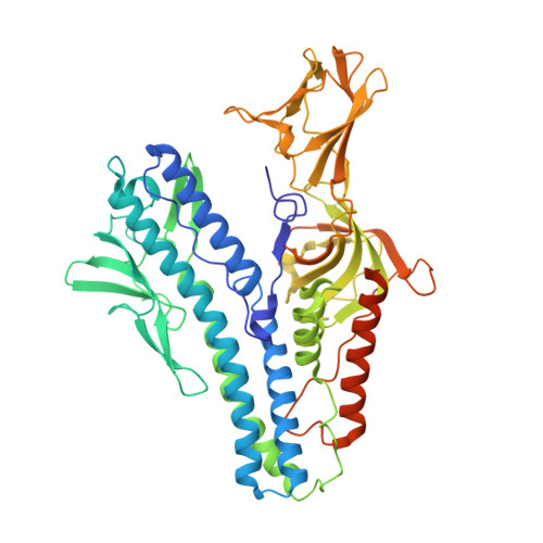
    Lipid membrane fusion is an essential function in many biological processes. Detailed mechanisms of membrane fusion and the protein structures involved have been mainly studied in eukaryotic systems, whereas very little is known about membrane fusion in prokaryotes. Haloarchaeal pleomorphic viruses (HRPVs) have a membrane envelope decorated with spikes that are presumed to be responsible for host attachment and membrane fusion. Here we determine atomic structures of the ectodomains of the 57-kDa spike protein VP5 from two related HRPVs revealing a previously unreported V-shaped fold. By Volta phase plate cryo-electron tomography we show that VP5 is monomeric on the viral surface, and we establish the orientation of the molecules with respect to the viral membrane. We also show that the viral membrane fuses with the host cytoplasmic membrane in a process mediated by VP5. This sheds light on protein structures involved in prokaryotic membrane fusion.


  • Organizational Affiliation

    Division of Structural Biology, Wellcome Centre for Human Genetics, University of Oxford, Oxford, OX3 7BN, UK.


Macromolecules
Find similar proteins by:  (by identity cutoff)  |  3D Structure
Entity ID: 1
MoleculeChains Sequence LengthOrganismDetailsImage
VP5533Halorubrum pleomorphic virus 2Mutation(s): 0 
UniProt
Find proteins for H9ABL9 (Halorubrum pleomorphic virus 2)
Explore H9ABL9 
Go to UniProtKB:  H9ABL9
Entity Groups  
Sequence Clusters30% Identity50% Identity70% Identity90% Identity95% Identity100% Identity
UniProt GroupH9ABL9
Sequence Annotations
Expand
  • Reference Sequence
Experimental Data & Validation

Experimental Data

  • Method: X-RAY DIFFRACTION
  • Resolution: 2.46 Å
  • R-Value Free: 0.241 
  • R-Value Work: 0.224 
  • R-Value Observed: 0.225 
  • Space Group: P 21 21 21
Unit Cell:
Length ( Å )Angle ( ˚ )
a = 48.08α = 90
b = 93.22β = 90
c = 121.82γ = 90
Software Package:
Software NamePurpose
BUSTERrefinement
xia2data reduction
xia2data scaling
PHASERphasing

Structure Validation

View Full Validation Report


Ligand Structure Quality Assessment 


Entry History & Funding Information

Deposition Data


Funding OrganizationLocationGrant Number
Medical Research Council (United Kingdom)United KingdomG1000099
Medical Research Council (United Kingdom)United KingdomMR/N00065X/1
Wellcome TrustUnited Kingdom090532/Z/09/Z
European Research CouncilUnited Kingdom649053
Academy of FinlandFinland283072
Academy of FinlandFinland255342

Revision History  (Full details and data files)

  • Version 1.0: 2019-02-27
    Type: Initial release
  • Version 1.1: 2020-07-29
    Type: Remediation
    Reason: Carbohydrate remediation
    Changes: Data collection, Derived calculations, Structure summary