6ONJ

Crystal structure of PPARgamma ligand binding domain in complex with TRAP220 peptide and agonist rosiglitazone


Experimental Data Snapshot

  • Method: X-RAY DIFFRACTION
  • Resolution: 2.30 Å
  • R-Value Free: 0.273 
  • R-Value Work: 0.212 
  • R-Value Observed: 0.218 

wwPDB Validation   3D Report Full Report


Ligand Structure Quality Assessment 


This is version 1.0 of the entry. See complete history


Literature

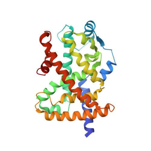
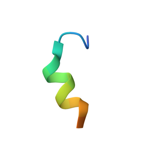
A molecular switch regulating transcriptional repression and activation of PPAR gamma.

Shang, J.Mosure, S.A.Zheng, J.Brust, R.Bass, J.Nichols, A.Solt, L.A.Griffin, P.R.Kojetin, D.J.

(2020) Nat Commun 11: 956-956

  • DOI: https://doi.org/10.1038/s41467-020-14750-x
  • Primary Citation of Related Structures:  
    6ONI, 6ONJ, 6PDZ

  • PubMed Abstract: 
  • Nuclear receptor (NR) transcription factors use a conserved activation function-2 (AF-2) helix 12 mechanism for agonist-induced coactivator interaction and NR transcriptional activation. In contrast, ligand-induced corepressor-dependent NR repression appears to occur through structurally diverse mechanisms ...

    Nuclear receptor (NR) transcription factors use a conserved activation function-2 (AF-2) helix 12 mechanism for agonist-induced coactivator interaction and NR transcriptional activation. In contrast, ligand-induced corepressor-dependent NR repression appears to occur through structurally diverse mechanisms. We report two crystal structures of peroxisome proliferator-activated receptor gamma (PPARγ) in an inverse agonist/corepressor-bound transcriptionally repressive conformation. Helix 12 is displaced from the solvent-exposed active conformation and occupies the orthosteric ligand-binding pocket enabled by a conformational change that doubles the pocket volume. Paramagnetic relaxation enhancement (PRE) NMR and chemical crosslinking mass spectrometry confirm the repressive helix 12 conformation. PRE NMR also defines the mechanism of action of the corepressor-selective inverse agonist T0070907, and reveals that apo-helix 12 exchanges between transcriptionally active and repressive conformations-supporting a fundamental hypothesis in the NR field that helix 12 exchanges between transcriptionally active and repressive conformations.


    Organizational Affiliation

    Department of Molecular Medicine, The Scripps Research Institute, Jupiter, FL, 33458, USA. dkojetin@scripps.edu.


Macromolecules
Find similar proteins by:  (by identity cutoff)  |  3D Structure
Entity ID: 1
MoleculeChainsSequence LengthOrganismDetailsImage
Peroxisome proliferator-activated receptor gammaA275Homo sapiensMutation(s): 0 
Gene Names: PPARGNR1C3
UniProt & NIH Common Fund Data Resources
Find proteins for P37231 (Homo sapiens)
Explore P37231 
Go to UniProtKB:  P37231
PHAROS:  P37231
Entity Groups  
Sequence Clusters30% Identity50% Identity70% Identity90% Identity95% Identity100% Identity
UniProt GroupP37231
Protein Feature View
Expand
  • Reference Sequence

Find similar proteins by:  Sequence   |   3D Structure  

Entity ID: 2
MoleculeChainsSequence LengthOrganismDetailsImage
Mediator of RNA polymerase II transcription subunit 1, TRAP220 Coactivator PeptideB [auth C]19Homo sapiensMutation(s): 0 
Gene Names: 
UniProt & NIH Common Fund Data Resources
Find proteins for Q15648 (Homo sapiens)
Explore Q15648 
Go to UniProtKB:  Q15648
PHAROS:  Q15648
Entity Groups  
Sequence Clusters30% Identity50% Identity70% Identity90% Identity95% Identity100% Identity
UniProt GroupQ15648
Protein Feature View
Expand
  • Reference Sequence
Small Molecules
Ligands 1 Unique
IDChainsName / Formula / InChI Key2D Diagram3D Interactions
BRL (Subject of Investigation/LOI)
Query on BRL

Download Ideal Coordinates CCD File 
C [auth A]2,4-THIAZOLIDIINEDIONE, 5-[[4-[2-(METHYL-2-PYRIDINYLAMINO)ETHOXY]PHENYL]METHYL]-(9CL)
C18 H19 N3 O3 S
YASAKCUCGLMORW-HNNXBMFYSA-N
 Ligand Interaction
Binding Affinity Annotations 
IDSourceBinding Affinity
BRL BindingDB:  6ONJ Ki: min: 8, max: 59 (nM) from 5 assay(s)
Kd: min: 0.14, max: 48 (nM) from 3 assay(s)
IC50: min: 1, max: 298 (nM) from 10 assay(s)
EC50: min: 1, max: 1600 (nM) from 35 assay(s)
Experimental Data & Validation

Experimental Data

  • Method: X-RAY DIFFRACTION
  • Resolution: 2.30 Å
  • R-Value Free: 0.273 
  • R-Value Work: 0.212 
  • R-Value Observed: 0.218 
  • Space Group: C 2 2 21
Unit Cell:
Length ( Å )Angle ( ˚ )
a = 54.386α = 90
b = 85.574β = 90
c = 121.629γ = 90
Software Package:
Software NamePurpose
PHENIXrefinement
SCALAdata scaling
PHASERphasing
PDB_EXTRACTdata extraction
DIALSdata reduction

Structure Validation

View Full Validation Report


Ligand Structure Quality Assessment 


Entry History & Funding Information

Deposition Data


Funding OrganizationLocationGrant Number
National Institutes of Health/National Institute of Diabetes and Digestive and Kidney Disease (NIH/NIDDK)United StatesR01DK101871

Revision History  (Full details and data files)

  • Version 1.0: 2020-03-04
    Type: Initial release