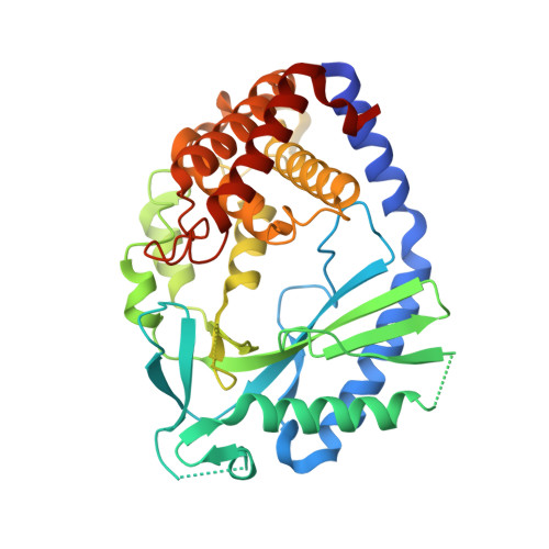
6O47

human cGAS core domain (K427E/K428E) bound with RU-521


Experimental Data Snapshot

  • Method: X-RAY DIFFRACTION
  • Resolution: 2.20 Å
  • R-Value Free: 0.221 
  • R-Value Work: 0.181 
  • R-Value Observed: 0.185 

wwPDB Validation   3D Report Full Report

Currently 6O47 does not have a validation slider image.


This is version 1.3 of the entry. See complete history


Literature

Human cGAS catalytic domain has an additional DNA-binding interface that enhances enzymatic activity and liquid-phase condensation.

Xie, W.Lama, L.Adura, C.Tomita, D.Glickman, J.F.Tuschl, T.Patel, D.J.

(2019) Proc Natl Acad Sci U S A 116: 11946-11955

  • DOI: https://doi.org/10.1073/pnas.1905013116
  • Primary Citation of Related Structures:  
    6EDB, 6EDC, 6O47

  • PubMed Abstract: 

    The cyclic GMP-AMP synthase (cGAS)-cGAMP-STING pathway plays a key role in innate immunity, with cGAS sensing both pathogenic and mislocalized DNA in the cytoplasm. Human cGAS (h-cGAS) constitutes an important drug target for control of antiinflammatory responses that can contribute to the onset of autoimmune diseases. Recent studies have established that the positively charged N-terminal segment of cGAS contributes to enhancement of cGAS enzymatic activity as a result of DNA-induced liquid-phase condensation. We have identified an additional cGAS CD -DNA interface (labeled site-C; CD, catalytic domain) in the crystal structure of a human SRY.cGAS CD -DNA complex, with mutations along this basic site-C cGAS interface disrupting liquid-phase condensation, as monitored by cGAMP formation, gel shift, spin-down, and turbidity assays, as well as time-lapse imaging of liquid droplet formation. We expand on an earlier ladder model of cGAS dimers bound to a pair of parallel-aligned DNAs to propose a multivalent interaction-mediated cluster model to account for DNA-mediated condensation involving both the N-terminal domain of cGAS and the site-C cGAS-DNA interface. We also report the crystal structure of the h-cGAS CD -DNA complex containing a triple mutant that disrupts the site-C interface, with this complex serving as a future platform for guiding cGAS inhibitor development at the DNA-bound h-cGAS level. Finally, we solved the structure of RU.521 bound in two alternate alignments to apo h-cGAS CD , thereby occupying more of the catalytic pocket and providing insights into further optimization of active-site-binding inhibitors.


  • Organizational Affiliation

    Structural Biology Program, Memorial Sloan Kettering Cancer Center, New York, NY 10065; xiew@mskcc.org pateld@mskcc.org.


Macromolecules
Find similar proteins by:  (by identity cutoff)  |  3D Structure
Entity ID: 1
MoleculeChains Sequence LengthOrganismDetailsImage
Cyclic GMP-AMP synthase372Homo sapiensMutation(s): 2 
Gene Names: CGASC6orf150MB21D1
EC: 2.7.7.86
UniProt & NIH Common Fund Data Resources
Find proteins for Q8N884 (Homo sapiens)
Explore Q8N884 
Go to UniProtKB:  Q8N884
PHAROS:  Q8N884
GTEx:  ENSG00000164430 
Entity Groups  
Sequence Clusters30% Identity50% Identity70% Identity90% Identity95% Identity100% Identity
UniProt GroupQ8N884
Sequence Annotations
Expand
  • Reference Sequence
Small Molecules
Ligands 4 Unique
IDChains Name / Formula / InChI Key2D Diagram3D Interactions
AEV
Query on AEV

Download Ideal Coordinates CCD File 
F [auth A]2-(4,5-dichloro-1H-benzimidazol-2-yl)-5-methyl-4-[(1R)-3-oxo-1,3-dihydro-2-benzofuran-1-yl]-1,2-dihydro-3H-pyrazol-3-one
C19 H12 Cl2 N4 O3
IAWZUQAOMURCLV-MRXNPFEDSA-N
LLS
Query on LLS

Download Ideal Coordinates CCD File 
E [auth A](3~{S})-3-[1-[4,5-bis(chloranyl)-1~{H}-benzimidazol-2-yl]-3-methyl-5-oxidanyl-pyrazol-4-yl]-3~{H}-2-benzofuran-1-one
C19 H12 Cl2 N4 O3
VIQXILLOJLATEF-INIZCTEOSA-N
CIT
Query on CIT

Download Ideal Coordinates CCD File 
B [auth A],
C [auth A]
CITRIC ACID
C6 H8 O7
KRKNYBCHXYNGOX-UHFFFAOYSA-N
ZN
Query on ZN

Download Ideal Coordinates CCD File 
D [auth A]ZINC ION
Zn
PTFCDOFLOPIGGS-UHFFFAOYSA-N
Experimental Data & Validation

Experimental Data

  • Method: X-RAY DIFFRACTION
  • Resolution: 2.20 Å
  • R-Value Free: 0.221 
  • R-Value Work: 0.181 
  • R-Value Observed: 0.185 
  • Space Group: P 64
Unit Cell:
Length ( Å )Angle ( ˚ )
a = 116.157α = 90
b = 116.157β = 90
c = 59.951γ = 120
Software Package:
Software NamePurpose
PHENIXrefinement
HKL-3000data reduction
HKL-3000data scaling
PHASERphasing

Structure Validation

View Full Validation Report

Currently 6O47 does not have a validation slider image.


Entry History 

Deposition Data

Revision History  (Full details and data files)

  • Version 1.0: 2019-05-29
    Type: Initial release
  • Version 1.1: 2019-06-12
    Changes: Data collection, Database references
  • Version 1.2: 2019-06-26
    Changes: Data collection, Database references
  • Version 1.3: 2023-10-11
    Changes: Data collection, Database references, Refinement description