6MAC

Ternary structure of GDF11 bound to ActRIIB-ECD and Alk5-ECD


Experimental Data Snapshot

  • Method: X-RAY DIFFRACTION
  • Resolution: 2.34 Å
  • R-Value Free: 0.267 
  • R-Value Work: 0.222 
  • R-Value Observed: 0.226 

wwPDB Validation   3D Report Full Report


Ligand Structure Quality Assessment 


This is version 1.4 of the entry. See complete history


Literature

Structural characterization of an activin class ternary receptor complex reveals a third paradigm for receptor specificity.

Goebel, E.J.Corpina, R.A.Hinck, C.S.Czepnik, M.Castonguay, R.Grenha, R.Boisvert, A.Miklossy, G.Fullerton, P.T.Matzuk, M.M.Idone, V.J.Economides, A.N.Kumar, R.Hinck, A.P.Thompson, T.B.

(2019) Proc Natl Acad Sci U S A 116: 15505-15513

  • DOI: https://doi.org/10.1073/pnas.1906253116
  • Primary Citation of Related Structures:  
    6MAC

  • PubMed Abstract: 

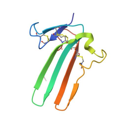
    TGFβ family ligands, which include the TGFβs, BMPs, and activins, signal by forming a ternary complex with type I and type II receptors. For TGFβs and BMPs, structures of ternary complexes have revealed differences in receptor assembly. However, structural information for how activins assemble a ternary receptor complex is lacking. We report the structure of an activin class member, GDF11, in complex with the type II receptor ActRIIB and the type I receptor Alk5. The structure reveals that receptor positioning is similar to the BMP class, with no interreceptor contacts; however, the type I receptor interactions are shifted toward the ligand fingertips and away from the dimer interface. Mutational analysis shows that ligand type I specificity is derived from differences in the fingertips of the ligands that interact with an extended loop specific to Alk4 and Alk5. The study also reveals differences for how TGFβ and GDF11 bind to the same type I receptor, Alk5. For GDF11, additional contacts at the fingertip region substitute for the interreceptor interactions that are seen for TGFβ, indicating that Alk5 binding to GDF11 is more dependent on direct contacts. In support, we show that a single residue of Alk5 (Phe 84 ), when mutated, abolishes GDF11 signaling, but has little impact on TGFβ signaling. The structure of GDF11/ActRIIB/Alk5 shows that, across the TGFβ family, different mechanisms regulate type I receptor binding and specificity, providing a molecular explanation for how the activin class accommodates low-affinity type I interactions without the requirement of cooperative receptor interactions.


  • Organizational Affiliation

    Department of Molecular Genetics, Biochemistry, and Microbiology, University of Cincinnati, Cincinnati, OH 45267.


Macromolecules
Find similar proteins by:  (by identity cutoff)  |  3D Structure
Entity ID: 1
MoleculeChains Sequence LengthOrganismDetailsImage
Growth/differentiation factor 11108Homo sapiensMutation(s): 0 
Gene Names: GDF11BMP11
UniProt & NIH Common Fund Data Resources
Find proteins for O95390 (Homo sapiens)
Explore O95390 
Go to UniProtKB:  O95390
PHAROS:  O95390
GTEx:  ENSG00000135414 
Entity Groups  
Sequence Clusters30% Identity50% Identity70% Identity90% Identity95% Identity100% Identity
UniProt GroupO95390
Sequence Annotations
Expand
  • Reference Sequence
Find similar proteins by:  (by identity cutoff)  |  3D Structure
Entity ID: 2
MoleculeChains Sequence LengthOrganismDetailsImage
Activin receptor type-2BB [auth C]95Rattus norvegicusMutation(s): 0 
Gene Names: Acvr2bActriib
EC: 2.7.11.30
UniProt
Find proteins for P38445 (Rattus norvegicus)
Explore P38445 
Go to UniProtKB:  P38445
Entity Groups  
Sequence Clusters30% Identity50% Identity70% Identity90% Identity95% Identity100% Identity
UniProt GroupP38445
Sequence Annotations
Expand
  • Reference Sequence
Find similar proteins by:  (by identity cutoff)  |  3D Structure
Entity ID: 3
MoleculeChains Sequence LengthOrganismDetailsImage
TGF-beta receptor type-1C [auth K]80Homo sapiensMutation(s): 0 
Gene Names: TGFBR1ALK5SKR4
EC: 2.7.11.30
UniProt & NIH Common Fund Data Resources
Find proteins for P36897 (Homo sapiens)
Explore P36897 
Go to UniProtKB:  P36897
PHAROS:  P36897
GTEx:  ENSG00000106799 
Entity Groups  
Sequence Clusters30% Identity50% Identity70% Identity90% Identity95% Identity100% Identity
UniProt GroupP36897
Sequence Annotations
Expand
  • Reference Sequence
Small Molecules
Ligands 1 Unique
IDChains Name / Formula / InChI Key2D Diagram3D Interactions
NAG
Query on NAG

Download Ideal Coordinates CCD File 
D [auth C],
E [auth C]
2-acetamido-2-deoxy-beta-D-glucopyranose
C8 H15 N O6
OVRNDRQMDRJTHS-FMDGEEDCSA-N
Experimental Data & Validation

Experimental Data

  • Method: X-RAY DIFFRACTION
  • Resolution: 2.34 Å
  • R-Value Free: 0.267 
  • R-Value Work: 0.222 
  • R-Value Observed: 0.226 
  • Space Group: P 32 1 2
Unit Cell:
Length ( Å )Angle ( ˚ )
a = 116.624α = 90
b = 116.624β = 90
c = 56.625γ = 120
Software Package:
Software NamePurpose
PHENIXrefinement
PDB_EXTRACTdata extraction
DIALSdata reduction
Aimlessdata scaling
PHASERphasing

Structure Validation

View Full Validation Report


Ligand Structure Quality Assessment 


Entry History 

Deposition Data

Revision History  (Full details and data files)

  • Version 1.0: 2019-07-17
    Type: Initial release
  • Version 1.1: 2019-07-31
    Changes: Data collection, Database references
  • Version 1.2: 2019-08-14
    Changes: Data collection, Database references
  • Version 1.3: 2020-07-29
    Type: Remediation
    Reason: Carbohydrate remediation
    Changes: Data collection, Derived calculations, Structure summary
  • Version 1.4: 2023-10-11
    Changes: Data collection, Database references, Refinement description, Structure summary