6KCX

Crystal structure of citrate complex of alpha-glucuronidase (TM0752)from Thermotoga maritima


Experimental Data Snapshot

  • Method: X-RAY DIFFRACTION
  • Resolution: 1.93 Å
  • R-Value Free: 0.184 
  • R-Value Work: 0.141 
  • R-Value Observed: 0.143 

wwPDB Validation   3D Report Full Report


Ligand Structure Quality Assessment 


This is version 1.0 of the entry. See complete history


Literature

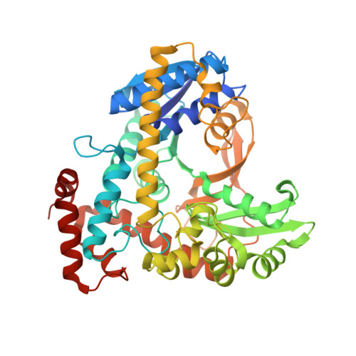
Structure of an alpha-glucuronidase in complex with Co2+and citrate provides insights into the mechanism and substrate recognition in the family 4 glycosyl hydrolases.

Mohapatra, S.B.Manoj, N.

(2019) Biochem Biophys Res Commun 518: 197-203

  • DOI: https://doi.org/10.1016/j.bbrc.2019.08.030
  • Primary Citation of Related Structures:  
    6KCX

  • PubMed Abstract: 
  • Glycosyl hydrolases belonging to the family 4 (GH4) use a unique redox-based NAD + -dependent reaction mechanism involving anionic intermediates and requires a divalent metal ion and reducing conditions for catalytic activity. These enzymes display wide specificity and selectivity for their substrates ...

    Glycosyl hydrolases belonging to the family 4 (GH4) use a unique redox-based NAD + -dependent reaction mechanism involving anionic intermediates and requires a divalent metal ion and reducing conditions for catalytic activity. These enzymes display wide specificity and selectivity for their substrates. However, the structural basis of substrate binding, recognition and specificity remains poorly studied. Here, we report the crystal structure of Thermotoga maritima TmAgu4B, a GH4 α-glucuronidase, in complex with Co 2+ and citrate. Analysis of GH4 structures show that the metal ion is present in a conserved octahedral coordination with conserved side chain atoms, the ligand atoms and an invariant water molecule. The data provides the first structural evidence for a metal-activated hydroxide ion that acts as the general base to deprotonate the C3-hydroxyl group of the glycone, a rate-limiting step in the mechanism. Furthermore, the citrate binding mode in the active site is analogous to a bound glucuronide substrate and provides insights into the mode of substrate interaction with the metal ion, the active site residues and, the structural basis of substrate recognition in a GH4 α-glucuronidase.


    Organizational Affiliation

    Department of Biotechnology, Bhupat and Jyoti Mehta School of Biosciences, Indian Institute of Technology Madras, Chennai, 600036, India. Electronic address: nmanoj@iitm.ac.in.


Macromolecules
Find similar proteins by:  (by identity cutoff)  |  3D Structure
Entity ID: 1
MoleculeChainsSequence LengthOrganismDetailsImage
Alpha-glucosidase, putativeA483Thermotoga maritima MSB8Mutation(s): 0 
Gene Names: TM_0752
UniProt
Find proteins for Q9WZL1 (Thermotoga maritima (strain ATCC 43589 / DSM 3109 / JCM 10099 / NBRC 100826 / MSB8))
Explore Q9WZL1 
Go to UniProtKB:  Q9WZL1
Entity Groups  
Sequence Clusters30% Identity50% Identity70% Identity90% Identity95% Identity100% Identity
UniProt GroupQ9WZL1
Protein Feature View
Expand
  • Reference Sequence
Small Molecules
Ligands 4 Unique
IDChainsName / Formula / InChI Key2D Diagram3D Interactions
CIT (Subject of Investigation/LOI)
Query on CIT

Download Ideal Coordinates CCD File 
E [auth A],
F [auth A]
CITRIC ACID
C6 H8 O7
KRKNYBCHXYNGOX-UHFFFAOYSA-N
 Ligand Interaction
IMD
Query on IMD

Download Ideal Coordinates CCD File 
C [auth A]IMIDAZOLE
C3 H5 N2
RAXXELZNTBOGNW-UHFFFAOYSA-O
 Ligand Interaction
IPA
Query on IPA

Download Ideal Coordinates CCD File 
D [auth A]ISOPROPYL ALCOHOL
C3 H8 O
KFZMGEQAYNKOFK-UHFFFAOYSA-N
 Ligand Interaction
CO (Subject of Investigation/LOI)
Query on CO

Download Ideal Coordinates CCD File 
B [auth A]COBALT (II) ION
Co
XLJKHNWPARRRJB-UHFFFAOYSA-N
 Ligand Interaction
Binding Affinity Annotations 
IDSourceBinding Affinity
CIT Binding MOAD:  6KCX IC50: 6.00e+6 (nM) from 1 assay(s)
Experimental Data & Validation

Experimental Data

  • Method: X-RAY DIFFRACTION
  • Resolution: 1.93 Å
  • R-Value Free: 0.184 
  • R-Value Work: 0.141 
  • R-Value Observed: 0.143 
  • Space Group: C 1 2 1
Unit Cell:
Length ( Å )Angle ( ˚ )
a = 74.42α = 90
b = 80.41β = 102.1
c = 89.05γ = 90
Software Package:
Software NamePurpose
PHENIXrefinement
MOSFLMdata reduction
Aimlessdata scaling
PHASERphasing
PDB_EXTRACTdata extraction

Structure Validation

View Full Validation Report


Ligand Structure Quality Assessment 


Entry History 

Deposition Data

Revision History  (Full details and data files)

  • Version 1.0: 2020-04-29
    Type: Initial release