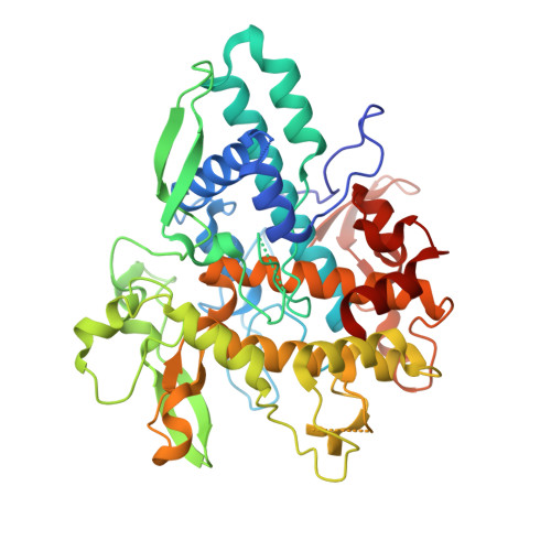
6JU9

Aspergillus oryzae active-tyrosinase copper-bound C92A mutant complexed with L-tyrosine


Experimental Data Snapshot

  • Method: X-RAY DIFFRACTION
  • Resolution: 1.42 Å
  • R-Value Free: 0.197 
  • R-Value Work: 0.154 
  • R-Value Observed: 0.156 

wwPDB Validation   3D Report Full Report


Ligand Structure Quality Assessment 


This is version 1.2 of the entry. See complete history


Literature

Copper-Oxygen Dynamics in the Tyrosinase Mechanism.

Fujieda, N.Umakoshi, K.Ochi, Y.Nishikawa, Y.Yanagisawa, S.Kubo, M.Kurisu, G.Itoh, S.

(2020) Angew Chem Int Ed Engl 59: 13385-13390

  • DOI: https://doi.org/10.1002/anie.202004733
  • Primary Citation of Related Structures:  
    6JU4, 6JU5, 6JU6, 6JU7, 6JU8, 6JU9, 6JUA, 6JUB, 6JUC, 6JUD

  • PubMed Abstract: 

    The dinuclear copper enzyme, tyrosinase, activates O 2 to form a (μ-η 2 2 -peroxido)dicopper(II) species, which hydroxylates phenols to catechols. However, the exact mechanism of phenolase reaction in the catalytic site of tyrosinase is still under debate. We herein report the near atomic resolution X-ray crystal structures of the active tyrosinases with substrate l-tyrosine. At their catalytic sites, CuA moved toward l-tyrosine (CuA1 → CuA2), whose phenol oxygen directly coordinates to CuA2, involving the movement of CuB (CuB1 → CuB2). The crystal structures and spectroscopic analyses of the dioxygen-bound tyrosinases demonstrated that the peroxide ligand rotated, spontaneously weakening its O-O bond. Thus, the copper migration induced by the substrate-binding is accompanied by rearrangement of the bound peroxide species so as to provide one of the peroxide oxygen atoms with access to the phenol substrate's ϵ carbon atom.


  • Organizational Affiliation

    Department of Applied Life Sciences, Graduate School of Life and Environmental Sciences, Osaka Prefecture University, 1-1 Gakuen-cho, Naka-ku, Sakai-shi, Osaka, 599-8531, Japan.


Macromolecules
Find similar proteins by:  (by identity cutoff)  |  3D Structure
Entity ID: 1
MoleculeChains Sequence LengthOrganismDetailsImage
Tyrosinase
A, B
461Aspergillus oryzaeMutation(s): 1 
Gene Names: OAory_01107480
EC: 1.14.18.1
UniProt
Find proteins for A0A1S9DK56 (Aspergillus oryzae)
Explore A0A1S9DK56 
Go to UniProtKB:  A0A1S9DK56
Entity Groups  
Sequence Clusters30% Identity50% Identity70% Identity90% Identity95% Identity100% Identity
UniProt GroupA0A1S9DK56
Sequence Annotations
Expand
  • Reference Sequence
Small Molecules
Ligands 4 Unique
IDChains Name / Formula / InChI Key2D Diagram3D Interactions
DAH
Query on DAH

Download Ideal Coordinates CCD File 
D [auth A]3,4-DIHYDROXYPHENYLALANINE
C9 H11 N O4
WTDRDQBEARUVNC-LURJTMIESA-N
TYR
Query on TYR

Download Ideal Coordinates CCD File 
C [auth A],
H [auth B]
TYROSINE
C9 H11 N O3
OUYCCCASQSFEME-QMMMGPOBSA-N
CU
Query on CU

Download Ideal Coordinates CCD File 
E [auth A],
F [auth A],
I [auth B],
J [auth B]
COPPER (II) ION
Cu
JPVYNHNXODAKFH-UHFFFAOYSA-N
NO3
Query on NO3

Download Ideal Coordinates CCD File 
G [auth A]NITRATE ION
N O3
NHNBFGGVMKEFGY-UHFFFAOYSA-N
Experimental Data & Validation

Experimental Data

  • Method: X-RAY DIFFRACTION
  • Resolution: 1.42 Å
  • R-Value Free: 0.197 
  • R-Value Work: 0.154 
  • R-Value Observed: 0.156 
  • Space Group: P 21 21 21
Unit Cell:
Length ( Å )Angle ( ˚ )
a = 54.544α = 90
b = 104.97β = 90
c = 154.877γ = 90
Software Package:
Software NamePurpose
DENZOdata reduction
HKL-2000data scaling
PHASERphasing
SHELXLrefinement

Structure Validation

View Full Validation Report


Ligand Structure Quality Assessment 


Entry History & Funding Information

Deposition Data


Funding OrganizationLocationGrant Number
Japan Science and TechnologyJapanCREST(JPMJCR16P1)

Revision History  (Full details and data files)

  • Version 1.0: 2020-05-13
    Type: Initial release
  • Version 1.1: 2020-08-12
    Changes: Database references, Derived calculations
  • Version 1.2: 2023-11-22
    Changes: Data collection, Database references, Refinement description