6IMV

The complex structure of endo-beta-1,2-glucanase from Talaromyces funiculosus with sophorose


Experimental Data Snapshot

  • Method: X-RAY DIFFRACTION
  • Resolution: 2.00 Å
  • R-Value Free: 0.209 
  • R-Value Work: 0.182 
  • R-Value Observed: 0.183 

wwPDB Validation   3D Report Full Report


Ligand Structure Quality Assessment 


This is version 2.1 of the entry. See complete history


Literature

Identification, characterization, and structural analyses of a fungal endo-beta-1,2-glucanase reveal a new glycoside hydrolase family.

Tanaka, N.Nakajima, M.Narukawa-Nara, M.Matsunaga, H.Kamisuki, S.Aramasa, H.Takahashi, Y.Sugimoto, N.Abe, K.Terada, T.Miyanaga, A.Yamashita, T.Sugawara, F.Kamakura, T.Komba, S.Nakai, H.Taguchi, H.

(2019) J Biol Chem 294: 7942-7965

  • DOI: https://doi.org/10.1074/jbc.RA118.007087
  • Primary Citation of Related Structures:  
    6IMU, 6IMV, 6IMW

  • PubMed Abstract: 

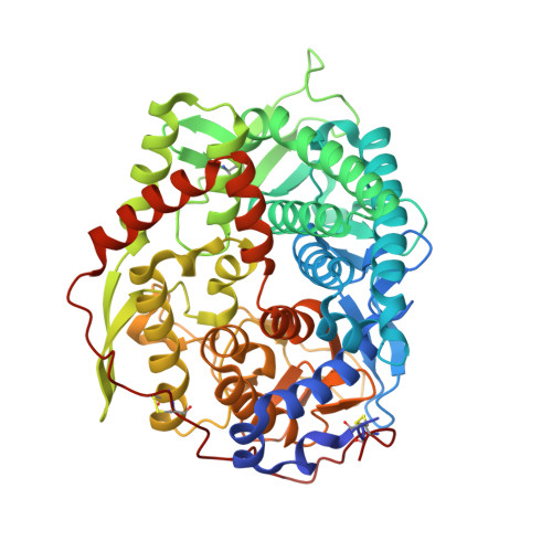
    endo -β-1,2-Glucanase (SGL) is an enzyme that hydrolyzes β-1,2-glucans, which play important physiological roles in some bacteria as a cyclic form. To date, no eukaryotic SGL has been identified. We purified an SGL from Talaromyces funiculosus ( Tf SGL), a soil fungus, to homogeneity and then cloned the complementary DNA encoding the enzyme. Tf SGL shows no significant sequence similarity to any known glycoside hydrolase (GH) families, but shows significant similarity to certain eukaryotic proteins with unknown functions. The recombinant Tf SGL ( Tf SGLr) specifically hydrolyzed linear and cyclic β-1,2-glucans to sophorose (Glc-β-1,2-Glc) as a main product. Tf SGLr hydrolyzed reducing-end-modified β-1,2-gluco-oligosaccharides to release a sophoroside with the modified moiety. These results indicate that Tf SGL is an endo -type enzyme that preferably releases sophorose from the reducing end of substrates. Stereochemical analysis demonstrated that Tf SGL is an inverting enzyme. The overall structure of Tf SGLr includes an (α/α) 6 toroid fold. The substrate-binding mode was revealed by the structure of a Michaelis complex of an inactive Tf SGLr mutant with a β-1,2-glucoheptasaccharide. Mutational analysis and action pattern analysis of β-1,2-gluco-oligosaccharide derivatives revealed an unprecedented catalytic mechanism for substrate hydrolysis. Glu-262 (general acid) indirectly protonates the anomeric oxygen at subsite -1 via the 3-hydroxy group of the Glc moiety at subsite +2, and Asp-446 (general base) activates the nucleophilic water via another water. Tf SGLr is apparently different from a GH144 SGL in the reaction and substrate recognition mechanism based on structural comparison. Overall, we propose that Tf SGL and closely-related enzymes can be classified into a new family, GH162.


  • Organizational Affiliation

    From the Department of Applied Biological Science, Faculty of Science and Technology, Tokyo University of Science, 2641 Yamazaki, Noda, Chiba 278-8510.


Macromolecules
Find similar proteins by:  (by identity cutoff)  |  3D Structure
Entity ID: 1
MoleculeChains Sequence LengthOrganismDetailsImage
Endo-beta-1,2-glucanase
A, B
505Talaromyces funiculosusMutation(s): 0 
EC: 3.2.1.71
UniProt
Find proteins for A0A4V8H012 (Talaromyces funiculosus)
Explore A0A4V8H012 
Go to UniProtKB:  A0A4V8H012
Entity Groups  
Sequence Clusters30% Identity50% Identity70% Identity90% Identity95% Identity100% Identity
UniProt GroupA0A4V8H012
Sequence Annotations
Expand
  • Reference Sequence
Oligosaccharides

Help

Entity ID: 2
MoleculeChains Length2D Diagram Glycosylation3D Interactions
2-acetamido-2-deoxy-beta-D-glucopyranose-(1-4)-2-acetamido-2-deoxy-beta-D-glucopyranose
C
2N-Glycosylation
Glycosylation Resources
GlyTouCan:  G42666HT
GlyCosmos:  G42666HT
GlyGen:  G42666HT
Entity ID: 3
MoleculeChains Length2D Diagram Glycosylation3D Interactions
beta-D-glucopyranose-(1-2)-alpha-D-glucopyranose
D, E
2N/A
Glycosylation Resources
GlyTouCan:  G71571NA
GlyCosmos:  G71571NA
Small Molecules
Ligands 3 Unique
IDChains Name / Formula / InChI Key2D Diagram3D Interactions
NAG (Subject of Investigation/LOI)
Query on NAG

Download Ideal Coordinates CCD File 
F [auth A]
G [auth A]
H [auth A]
L [auth B]
M [auth B]
F [auth A],
G [auth A],
H [auth A],
L [auth B],
M [auth B],
N [auth B],
O [auth B]
2-acetamido-2-deoxy-beta-D-glucopyranose
C8 H15 N O6
OVRNDRQMDRJTHS-FMDGEEDCSA-N
BGC (Subject of Investigation/LOI)
Query on BGC

Download Ideal Coordinates CCD File 
I [auth A],
P [auth B]
beta-D-glucopyranose
C6 H12 O6
WQZGKKKJIJFFOK-VFUOTHLCSA-N
PEG (Subject of Investigation/LOI)
Query on PEG

Download Ideal Coordinates CCD File 
J [auth A],
K [auth A],
Q [auth B]
DI(HYDROXYETHYL)ETHER
C4 H10 O3
MTHSVFCYNBDYFN-UHFFFAOYSA-N
Experimental Data & Validation

Experimental Data

  • Method: X-RAY DIFFRACTION
  • Resolution: 2.00 Å
  • R-Value Free: 0.209 
  • R-Value Work: 0.182 
  • R-Value Observed: 0.183 
  • Space Group: P 21 21 2
Unit Cell:
Length ( Å )Angle ( ˚ )
a = 92.088α = 90
b = 118.104β = 90
c = 93.428γ = 90
Software Package:
Software NamePurpose
REFMACrefinement
XDSdata reduction
Aimlessdata scaling
MOLREPphasing

Structure Validation

View Full Validation Report


Ligand Structure Quality Assessment 


Entry History 

Revision History  (Full details and data files)

  • Version 1.0: 2019-04-10
    Type: Initial release
  • Version 1.1: 2019-05-22
    Changes: Data collection, Database references
  • Version 2.0: 2020-07-29
    Type: Remediation
    Reason: Carbohydrate remediation
    Changes: Atomic model, Data collection, Derived calculations, Structure summary
  • Version 2.1: 2023-11-22
    Changes: Data collection, Database references, Derived calculations, Refinement description, Structure summary