6H6A | pdb_00006h6a

Crystal structure of UNC119 in complex with LCK peptide


Experimental Data Snapshot

  • Method: X-RAY DIFFRACTION
  • Resolution: 2.00 Å
  • R-Value Free: 
    0.212 (Depositor), 0.216 (DCC) 
  • R-Value Work: 
    0.171 (Depositor), 0.181 (DCC) 
  • R-Value Observed: 
    0.173 (Depositor) 

wwPDB Validation 3D Report Full Report

Validation slider image for 6H6A

Ligand Structure Quality Assessment 


This is version 1.4 of the entry. See complete history

Literature

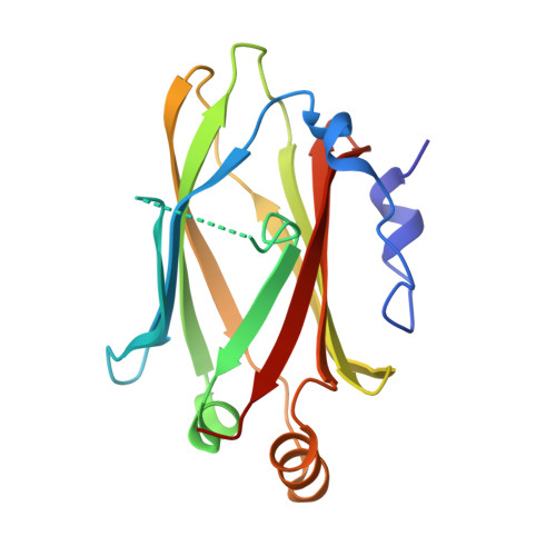
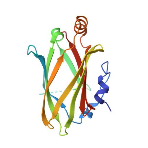
The Ciliary Machinery Is Repurposed for T Cell Immune Synapse Trafficking of LCK.

Stephen, L.A.ElMaghloob, Y.McIlwraith, M.J.Yelland, T.Castro Sanchez, P.Roda-Navarro, P.Ismail, S.

(2018) Dev Cell 47: 122-132.e4

  • DOI: https://doi.org/10.1016/j.devcel.2018.08.012
  • Primary Citation Related Structures: 
    6H6A

  • PubMed Abstract: 

    Upon engagement of the T cell receptor with an antigen-presenting cell, LCK initiates TCR signaling by phosphorylating its activation motifs. However, the mechanism of LCK activation specifically at the immune synapse is a major question. We show that phosphorylation of the LCK activating Y394, despite modestly increasing its catalytic rate, dramatically focuses LCK localization to the immune synapse. We describe a trafficking mechanism whereby UNC119A extracts membrane-bound LCK by sequestering the hydrophobic myristoyl group, followed by release at the target membrane under the control of the ciliary ARL3/ARL13B. The UNC119A N terminus acts as a "regulatory arm" by binding the LCK kinase domain, an interaction inhibited by LCK Y394 phosphorylation, thus together with the ARL3/ARL13B machinery ensuring immune synapse focusing of active LCK. We propose that the ciliary machinery has been repurposed by T cells to generate and maintain polarized segregation of signals such as activated LCK at the immune synapse.


  • Organizational Affiliation
    • CR-UK Beatson Institute, Garscube Estate, Switchback Road, Glasgow G61 1BD, UK.

Macromolecule Content 

  • Total Structure Weight: 70.25 kDa 
  • Atom Count: 4,672 
  • Modeled Residue Count: 513 
  • Deposited Residue Count: 576 
  • Unique protein chains: 3

Macromolecules

Find similar proteins by:|  3D Structure
Entity ID: 1
MoleculeChains  Sequence LengthOrganismDetailsImage
Protein unc-119 homolog AA [auth D]182Homo sapiensMutation(s): 0 
Gene Names: UNC119RG4
UniProt & NIH Common Fund Data Resources
Find proteins for Q13432 (Homo sapiens)
Explore Q13432 
Go to UniProtKB:  Q13432
PHAROS:  Q13432
GTEx:  ENSG00000109103 
Entity Groups
Sequence Clusters30% Identity50% Identity70% Identity90% Identity95% Identity100% Identity
UniProt GroupQ13432
Sequence Annotations
Expand
Reference Sequence
Find similar proteins by:  Sequence   |   3D Structure  
Entity ID: 2
MoleculeChains  Sequence LengthOrganismDetailsImage
GLY-CYS-GLY-CYS-SER-SERB [auth E],
D [auth H],
F [auth K]
10Homo sapiensMutation(s): 0 
EC: 2.7.10.2
UniProt & NIH Common Fund Data Resources
Find proteins for P06239 (Homo sapiens)
Explore P06239 
Go to UniProtKB:  P06239
PHAROS:  P06239
GTEx:  ENSG00000182866 
Entity Groups
Sequence Clusters30% Identity50% Identity70% Identity90% Identity95% Identity100% Identity
UniProt GroupP06239
Sequence Annotations
Expand
Reference Sequence
Find similar proteins by:|  3D Structure
Entity ID: 3
MoleculeChains  Sequence LengthOrganismDetailsImage
Protein unc-119 homolog AC [auth G],
E [auth J]
182Homo sapiensMutation(s): 0 
Gene Names: UNC119RG4
UniProt & NIH Common Fund Data Resources
Find proteins for Q13432 (Homo sapiens)
Explore Q13432 
Go to UniProtKB:  Q13432
PHAROS:  Q13432
GTEx:  ENSG00000109103 
Entity Groups
Sequence Clusters30% Identity50% Identity70% Identity90% Identity95% Identity100% Identity
UniProt GroupQ13432
Sequence Annotations
Expand
Reference Sequence

Small Molecules

Ligands 4 Unique
IDChains Name / Formula / InChI Key2D Diagram3D Interactions
15P

Query on 15P


Download:Ideal Coordinates CCD File
I [auth D]POLYETHYLENE GLYCOL (N=34)
C69 H140 O35
VUYXVWGKCKTUMF-UHFFFAOYSA-N
MYR

Query on MYR


Download:Ideal Coordinates CCD File
J [auth E],
N [auth H],
S [auth K]
MYRISTIC ACID
C14 H28 O2
TUNFSRHWOTWDNC-UHFFFAOYSA-N
PO4

Query on PO4


Download:Ideal Coordinates CCD File
M [auth G],
R [auth J]
PHOSPHATE ION
O4 P
NBIIXXVUZAFLBC-UHFFFAOYSA-K
GOL

Query on GOL


Download:Ideal Coordinates CCD File
G [auth D]
H [auth D]
K [auth G]
L [auth G]
O [auth J]
G [auth D],
H [auth D],
K [auth G],
L [auth G],
O [auth J],
P [auth J],
Q [auth J]
GLYCEROL
C3 H8 O3
PEDCQBHIVMGVHV-UHFFFAOYSA-N

Experimental Data & Validation

Experimental Data

  • Method: X-RAY DIFFRACTION
  • Resolution: 2.00 Å
  • R-Value Free:  0.212 (Depositor), 0.216 (DCC) 
  • R-Value Work:  0.171 (Depositor), 0.181 (DCC) 
  • R-Value Observed: 0.173 (Depositor) 
Space Group: P 21 21 21
Unit Cell:
Length ( Å )Angle ( ˚ )
a = 50.09α = 90
b = 63.62β = 90
c = 188.16γ = 90
Software Package:
Software NamePurpose
REFMACrefinement
XDSdata reduction
Aimlessdata scaling
MOLREPphasing

Structure Validation

View Full Validation Report


Ligand Structure Quality Assessment 


Entry History 

& Funding Information

Deposition Data


Funding OrganizationLocationGrant Number
Cancer Research UKUnited Kingdom--

Revision History  (Full details and data files)

  • Version 1.0: 2018-09-26
    Type: Initial release
  • Version 1.1: 2019-04-10
    Changes: Data collection, Database references
  • Version 1.2: 2019-10-16
    Changes: Data collection
  • Version 1.3: 2019-11-27
    Changes: Derived calculations
  • Version 1.4: 2024-10-23
    Changes: Data collection, Database references, Refinement description, Structure summary