Exploring the interactions between model proteins and Pd(ii) or Pt(ii) compounds bearing charged N,N-pyridylbenzimidazole bidentate ligands by X-ray crystallography.
Ferraro, G., Mansour, A.M., Merlino, A.(2018) Dalton Trans 47: 10130-10138
- PubMed: 30004541 Search on PubMed
- DOI: https://doi.org/10.1039/c8dt01663a
- Primary Citation Related Structures:
6GOB, 6GOH, 6GOI, 6GOJ, 6GOK - PubMed Abstract:
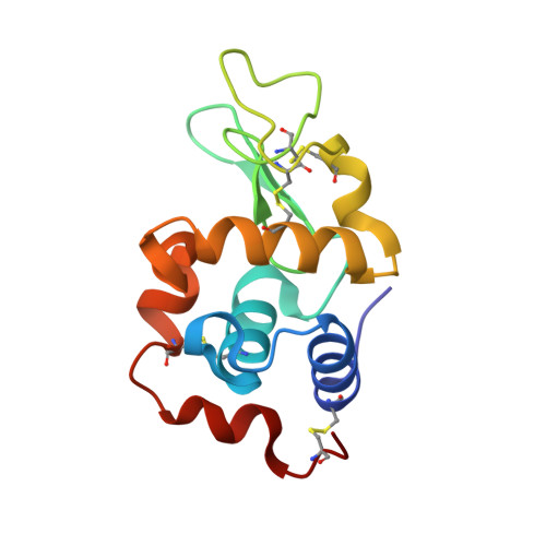
Pd(ii) and Pt(ii) compounds bearing N,N-pyridylbenzimidazole derivatives with an alkylated sulfonate or phosphonium side chain are able to bind the model protein lysozyme both covalently and non-covalently as an entire compound or as a product of a hydrolysis reaction. The interactions with the protein and the origin of the different behaviors of these complexes were unknown hitherto. Here, we present four crystal structures of their adducts with lysozyme. Pt- and Pd-containing fragments bind the protein with different stoichiometries close to the side chains of His15, Asp87, Asp101 and Asn77. The compounds bearing a phosphonium side chain degrade during the reaction with lysozyme. Data show the origin of the non-covalent mode of binding of Pd and Pt compounds bearing a sulfonate side chain, which drives the recognition process by forming a series of H-bonds and coulombic interactions with positively charged residue side chains. In a separate experiment, the structure of the adduct that is formed when the Pd(ii) compound containing an alkylated sulfonate group reacts with ribonuclease A was also determined. In this structure, the sulfonate-Pd(ii) complex binds the side chain of His105 on the surface of the protein and the side chain of the catalytically important His119 residue. Altogether, our data provide a structural basis for understanding the behavior of the analyzed Pd(ii)- and Pt(ii)-based cisplatin analogues in their reactions with proteins and show the first structural characterization of an adduct comprising a cisplatin analogue that is non-covalently bound to a protein. The results suggest that functionalization of a ligand system with a sulfonate group can significantly alter the protein-binding activity and thus the overall pharmacological profile of Pd(ii)- and Pt(ii)-based drugs.
- Department of Chemical Sciences, University of Naples Federico II, Complesso Universitario di Monte Sant'Angelo, 80126 Napoli, Italy. antonello.merlino@unina.it.
Organizational Affiliation: