6G85

Structure of Cdc14 bound to CBK1 PxL motif


Experimental Data Snapshot

  • Method: X-RAY DIFFRACTION
  • Resolution: 1.53 Å
  • R-Value Free: 0.209 
  • R-Value Work: 0.182 
  • R-Value Observed: 0.183 

wwPDB Validation   3D Report Full Report


This is version 1.3 of the entry. See complete history


Literature

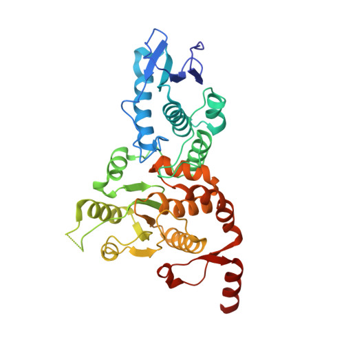
A PxL motif promotes timely cell cycle substrate dephosphorylation by the Cdc14 phosphatase.

Kataria, M.Mouilleron, S.Seo, M.H.Corbi-Verge, C.Kim, P.M.Uhlmann, F.

(2018) Nat Struct Mol Biol 25: 1093-1102

  • DOI: https://doi.org/10.1038/s41594-018-0152-3
  • Primary Citation of Related Structures:  
    6G84, 6G85, 6G86

  • PubMed Abstract: 

    The cell division cycle consists of a series of temporally ordered events. Cell cycle kinases and phosphatases provide key regulatory input, but how the correct substrate phosphorylation and dephosphorylation timing is achieved is incompletely understood. Here we identify a PxL substrate recognition motif that instructs dephosphorylation by the budding yeast Cdc14 phosphatase during mitotic exit. The PxL motif was prevalent in Cdc14-binding peptides enriched in a phage display screen of native disordered protein regions. PxL motif removal from the Cdc14 substrate Cbk1 delays its dephosphorylation, whereas addition of the motif advances dephosphorylation of otherwise late Cdc14 substrates. Crystal structures of Cdc14 bound to three PxL motif substrate peptides provide a molecular explanation for PxL motif recognition on the phosphatase surface. Our results illustrate the sophistication of phosphatase-substrate interactions and identify them as an important determinant of ordered cell cycle progression.


  • Organizational Affiliation

    Chromosome Segregation Laboratory, The Francis Crick Institute, London, UK.


Macromolecules
Find similar proteins by:  (by identity cutoff)  |  3D Structure
Entity ID: 1
MoleculeChains Sequence LengthOrganismDetailsImage
Tyrosine-protein phosphatase CDC14
A, B
374Saccharomyces cerevisiae S288CMutation(s): 0 
Gene Names: CDC14OAF3YFR028C
EC: 3.1.3.48
UniProt
Find proteins for Q00684 (Saccharomyces cerevisiae (strain ATCC 204508 / S288c))
Explore Q00684 
Go to UniProtKB:  Q00684
Entity Groups  
Sequence Clusters30% Identity50% Identity70% Identity90% Identity95% Identity100% Identity
UniProt GroupQ00684
Sequence Annotations
Expand
  • Reference Sequence

Find similar proteins by:  Sequence   |   3D Structure  

Entity ID: 2
MoleculeChains Sequence LengthOrganismDetailsImage
CBK116Saccharomyces cerevisiaeMutation(s): 0 
UniProt
Find proteins for P53894 (Saccharomyces cerevisiae (strain ATCC 204508 / S288c))
Explore P53894 
Go to UniProtKB:  P53894
Entity Groups  
Sequence Clusters30% Identity50% Identity70% Identity90% Identity95% Identity100% Identity
UniProt GroupP53894
Sequence Annotations
Expand
  • Reference Sequence

Find similar proteins by:  Sequence   |   3D Structure  

Entity ID: 3
MoleculeChains Sequence LengthOrganismDetailsImage
CBK117Saccharomyces cerevisiaeMutation(s): 0 
UniProt
Find proteins for P53894 (Saccharomyces cerevisiae (strain ATCC 204508 / S288c))
Explore P53894 
Go to UniProtKB:  P53894
Entity Groups  
Sequence Clusters30% Identity50% Identity70% Identity90% Identity95% Identity100% Identity
UniProt GroupP53894
Sequence Annotations
Expand
  • Reference Sequence
Small Molecules
Ligands 5 Unique
IDChains Name / Formula / InChI Key2D Diagram3D Interactions
MES
Query on MES

Download Ideal Coordinates CCD File 
E [auth A],
M [auth B]
2-(N-MORPHOLINO)-ETHANESULFONIC ACID
C6 H13 N O4 S
SXGZJKUKBWWHRA-UHFFFAOYSA-N
PEG
Query on PEG

Download Ideal Coordinates CCD File 
K [auth A]DI(HYDROXYETHYL)ETHER
C4 H10 O3
MTHSVFCYNBDYFN-UHFFFAOYSA-N
GOL
Query on GOL

Download Ideal Coordinates CCD File 
F [auth A]GLYCEROL
C3 H8 O3
PEDCQBHIVMGVHV-UHFFFAOYSA-N
ZN
Query on ZN

Download Ideal Coordinates CCD File 
L [auth A]ZINC ION
Zn
PTFCDOFLOPIGGS-UHFFFAOYSA-N
EDO
Query on EDO

Download Ideal Coordinates CCD File 
G [auth A],
H [auth A],
I [auth A],
J [auth A]
1,2-ETHANEDIOL
C2 H6 O2
LYCAIKOWRPUZTN-UHFFFAOYSA-N
Experimental Data & Validation

Experimental Data

  • Method: X-RAY DIFFRACTION
  • Resolution: 1.53 Å
  • R-Value Free: 0.209 
  • R-Value Work: 0.182 
  • R-Value Observed: 0.183 
  • Space Group: P 21 21 21
Unit Cell:
Length ( Å )Angle ( ˚ )
a = 76.154α = 90
b = 97.408β = 90
c = 129.176γ = 90
Software Package:
Software NamePurpose
PHENIXrefinement
xia2data reduction
Aimlessdata scaling
PHASERphasing

Structure Validation

View Full Validation Report


Entry History 

Deposition Data

Revision History  (Full details and data files)

  • Version 1.0: 2018-10-17
    Type: Initial release
  • Version 1.1: 2018-11-28
    Changes: Data collection, Database references, Structure summary
  • Version 1.2: 2018-12-19
    Changes: Data collection, Database references
  • Version 1.3: 2024-01-17
    Changes: Data collection, Database references, Refinement description