6F9Y | pdb_00006f9y

Lysozyme crystallized in presence of 10 mM lithium sulphate at pH 4.5

  • Classification: HYDROLASE
  • Organism(s): Gallus gallus
  • Mutation(s): No 

  • Deposited: 2017-12-15 Released: 2018-05-09 
  • Deposition Author(s): Camara-Artigas, A.
  • Funding Organization(s): Spanish Ministry of Economy and Competitiveness

Experimental Data Snapshot

  • Method: X-RAY DIFFRACTION
  • Resolution: 1.20 Å
  • R-Value Free: 
    0.201 (Depositor), 0.172 (DCC) 
  • R-Value Work: 
    0.167 (Depositor), 0.170 (DCC) 
  • R-Value Observed: 
    0.169 (Depositor) 

Starting Model: experimental
View more details

wwPDB Validation 3D Report Full Report

Validation slider image for 6F9Y

This is version 1.2 of the entry. See complete history

Literature

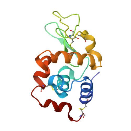
Orthorhombic lysozyme crystallization at acidic pH values driven by phosphate binding.

Plaza-Garrido, M.Salinas-Garcia, M.C.Camara-Artigas, A.

(2018) Acta Crystallogr D Struct Biol 74: 480-489

  • DOI: https://doi.org/10.1107/S205979831800517X
  • Primary Citation Related Structures: 
    6F1L, 6F1M, 6F1O, 6F1P, 6F1R, 6F9X, 6F9Y, 6F9Z, 6FA0

  • PubMed Abstract: 

    The structure of orthorhombic lysozyme has been obtained at 298 K and pH 4.5 using sodium chloride as the precipitant and in the presence of sodium phosphate at a concentration as low as 5 mM. Crystals belonging to space group P2 1 2 1 2 1 (unit-cell parameters a = 30, b = 56, c = 73 Å, α = β = γ = 90.00°) diffracted to a resolution higher than 1 Å, and the high quality of these crystals permitted the identification of a phosphate ion bound to Arg14 and His15. The binding of this ion produces long-range conformational changes affecting the loop containing Ser60-Asn74. The negatively charged phosphate ion shields the electrostatic repulsion of the positively charged arginine and histidine residues, resulting in higher stability of the phosphate-bound lysozyme. Additionally, a low-humidity orthorhombic variant was obtained at pH 4.5, and comparison with those previously obtained at pH 6.5 and 9.5 shows a 1.5 Å displacement of the fifth α-helix towards the active-site cavity, which might be relevant to protein function. Since lysozyme is broadly used as a model protein in studies related to protein crystallization and amyloid formation, these results indicate that the interaction of some anions must be considered when analysing experiments performed at acidic pH values.


  • Organizational Affiliation
    • Department of Chemistry and Physics, Agrifood Campus of International Excellence (ceiA3) and CIAMBITAL, University of Almería, Carretera de Sacramento, 04120 Almeria, Spain.

Macromolecule Content 

  • Total Structure Weight: 14.19 kDa 
  • Atom Count: 1,175 
  • Modeled Residue Count: 127 
  • Deposited Residue Count: 127 
  • Unique protein chains: 1

Macromolecules

Find similar proteins by:|  3D Structure
Entity ID: 1
MoleculeChains  Sequence LengthOrganismDetailsImage
Lysozyme C127Gallus gallusMutation(s): 0 
EC: 3.2.1.17
UniProt
Find proteins for P00698 (Gallus gallus)
Explore P00698 
Go to UniProtKB:  P00698
Entity Groups
Sequence Clusters30% Identity50% Identity70% Identity90% Identity95% Identity100% Identity
UniProt GroupP00698
Sequence Annotations
Expand
Reference Sequence

Experimental Data & Validation

Experimental Data

  • Method: X-RAY DIFFRACTION
  • Resolution: 1.20 Å
  • R-Value Free:  0.201 (Depositor), 0.172 (DCC) 
  • R-Value Work:  0.167 (Depositor), 0.170 (DCC) 
  • R-Value Observed: 0.169 (Depositor) 
Space Group: P 21 21 21
Unit Cell:
Length ( Å )Angle ( ˚ )
a = 30.131α = 90
b = 56.403β = 90
c = 73.429γ = 90
Software Package:
Software NamePurpose
XDSdata reduction
Aimlessdata scaling
PHASERphasing
PHENIXrefinement
PDB_EXTRACTdata extraction

Structure Validation

View Full Validation Report


Entry History 

& Funding Information

Deposition Data


Funding OrganizationLocationGrant Number
Spanish Ministry of Economy and CompetitivenessSpainBIO2016-78020-R

Revision History  (Full details and data files)

  • Version 1.0: 2018-05-09
    Type: Initial release
  • Version 1.1: 2024-01-17
    Changes: Data collection, Database references, Refinement description
  • Version 1.2: 2024-10-09
    Changes: Structure summary