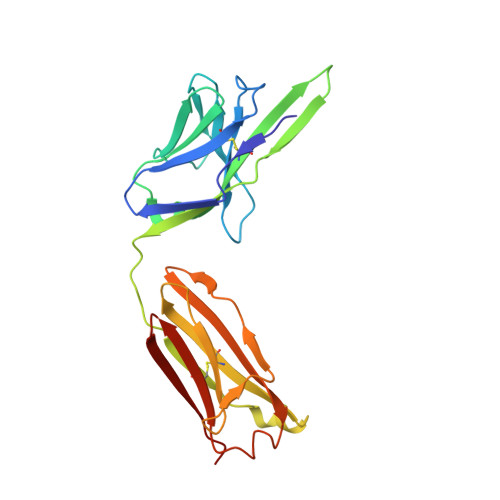
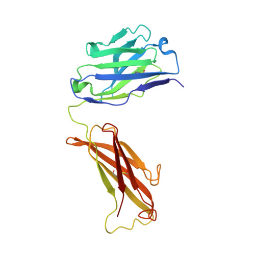
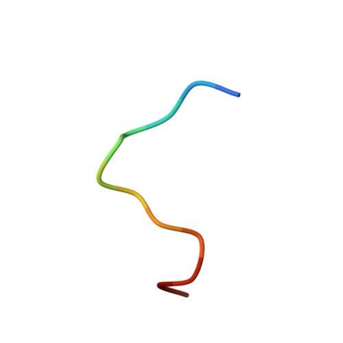
6F7T

Crystal Structure of an Fab fragment in complex with a peptide from Bacillus subtilis RNase Y


Experimental Data Snapshot

  • Method: X-RAY DIFFRACTION
  • Resolution: 2.60 Å
  • R-Value Free: 0.253 
  • R-Value Work: 0.202 
  • R-Value Observed: 0.204 

Starting Model: experimental
View more details

wwPDB Validation   3D Report Full Report


This is version 1.3 of the entry. See complete history


Literature

Dissociation of the Dimer of the Intrinsically Disordered Domain of RNase Y upon Antibody Binding.

Hardouin, P.Velours, C.Bou-Nader, C.Assrir, N.Laalami, S.Putzer, H.Durand, D.Golinelli-Pimpaneau, B.

(2018) Biophys J 115: 2102-2113

  • DOI: https://doi.org/10.1016/j.bpj.2018.10.016
  • Primary Citation of Related Structures:  
    6F7T

  • PubMed Abstract: 

    Although RNase Y acts as the key enzyme initiating messenger RNA decay in Bacillus subtilis and likely in many other Gram-positive bacteria, its three-dimensional structure remains unknown. An antibody belonging to the rare immunoglobulin G (IgG) 2b λx isotype was raised against a 12-residue conserved peptide from the N-terminal noncatalytic domain of B. subtilis RNase Y (BsRNaseY) that is predicted to be intrinsically disordered. Here, we show that this domain can be produced as a stand-alone protein called Nter-BsRNaseY that undergoes conformational changes between monomeric and dimeric forms. Circular dichroism and size exclusion chromatography coupled with multiangle light scattering or with small angle x-ray scattering indicate that the Nter-BsRNaseY dimer displays an elongated form and a high content of α-helices, in agreement with the existence of a central coiled-coil structure appended with flexible ends, and that the monomeric state of Nter-BsRNaseY is favored upon binding the fragment antigen binding (Fab) of the antibody. The dissociation constants of the IgG/BsRNaseY, IgG/Nter-BsRNaseY, and IgG/peptide complexes indicate that the affinity of the IgG for Nter-BsRNaseY is in the nM range and suggest that the peptide is less accessible in BsRNaseY than in Nter-BsRNaseY. The crystal structure of the Fab in complex with the peptide antigen shows that the peptide adopts an elongated U-shaped conformation in which the unique hydrophobic residue of the peptide, Leu6, is completely buried. The peptide/Fab complex may mimic the interaction of a microdomain of the N-terminal domain of BsRNaseY with one of its cellular partners within the degradosome complex. Altogether, our results suggest that BsRNaseY may become accessible for protein interaction upon dissociation of its N-terminal domain into the monomeric form.


  • Organizational Affiliation

    Laboratoire de Chimie des Processus Biologiques, UMR 8229 CNRS, Collège de France, Université Pierre et Marie Curie, Paris CEDEX 05, France.


Macromolecules
Find similar proteins by:  (by identity cutoff)  |  3D Structure
Entity ID: 1
MoleculeChains Sequence LengthOrganismDetailsImage
FAB RY79-90, LIGHT CHAINA,
F [auth L]
218Mus musculusMutation(s): 0 
Entity Groups  
Sequence Clusters30% Identity50% Identity70% Identity90% Identity95% Identity100% Identity
Sequence Annotations
Expand
  • Reference Sequence
Find similar proteins by:  (by identity cutoff)  |  3D Structure
Entity ID: 2
MoleculeChains Sequence LengthOrganismDetailsImage
FAB RY79-90, HEAVY CHAINB,
E [auth H]
211Mus musculusMutation(s): 0 
Entity Groups  
Sequence Clusters30% Identity50% Identity70% Identity90% Identity95% Identity100% Identity
Sequence Annotations
Expand
  • Reference Sequence

Find similar proteins by:  Sequence   |   3D Structure  

Entity ID: 3
MoleculeChains Sequence LengthOrganismDetailsImage
Ribonuclease Y
C, D
12Bacillus subtilisMutation(s): 0 
EC: 3.1
UniProt
Find proteins for O31774 (Bacillus subtilis (strain 168))
Explore O31774 
Go to UniProtKB:  O31774
Entity Groups  
Sequence Clusters30% Identity50% Identity70% Identity90% Identity95% Identity100% Identity
UniProt GroupO31774
Sequence Annotations
Expand
  • Reference Sequence
Small Molecules
Ligands 1 Unique
IDChains Name / Formula / InChI Key2D Diagram3D Interactions
MG
Query on MG

Download Ideal Coordinates CCD File 
G [auth L]MAGNESIUM ION
Mg
JLVVSXFLKOJNIY-UHFFFAOYSA-N
Experimental Data & Validation

Experimental Data

  • Method: X-RAY DIFFRACTION
  • Resolution: 2.60 Å
  • R-Value Free: 0.253 
  • R-Value Work: 0.202 
  • R-Value Observed: 0.204 
  • Space Group: P 21 21 2
Unit Cell:
Length ( Å )Angle ( ˚ )
a = 98.68α = 90
b = 130.45β = 90
c = 72.05γ = 90
Software Package:
Software NamePurpose
BUSTERrefinement
XDSdata reduction
XSCALEdata scaling
PHASERphasing

Structure Validation

View Full Validation Report


Entry History & Funding Information

Deposition Data


Funding OrganizationLocationGrant Number
LABEX DynamoFranceANR-11-LABX-0011
French National Research AgencyFranceANR-11-BSV8-026

Revision History  (Full details and data files)

  • Version 1.0: 2018-10-31
    Type: Initial release
  • Version 1.1: 2018-11-28
    Changes: Data collection, Database references
  • Version 1.2: 2018-12-19
    Changes: Data collection, Database references
  • Version 1.3: 2024-01-17
    Changes: Data collection, Database references, Refinement description