6D7K

Complex structure of Methane monooxygenase hydroxylase in complex with inhibitory subunit


Experimental Data Snapshot

  • Method: X-RAY DIFFRACTION
  • Resolution: 2.60 Å
  • R-Value Free: 0.221 
  • R-Value Work: 0.178 
  • R-Value Observed: 0.180 

wwPDB Validation   3D Report Full Report


This is version 1.4 of the entry. See complete history


Literature

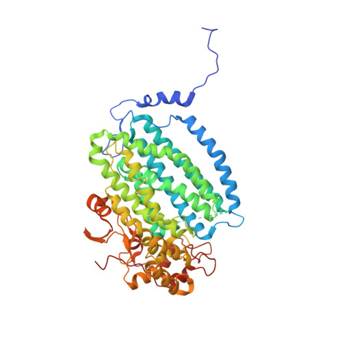
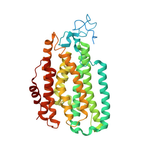
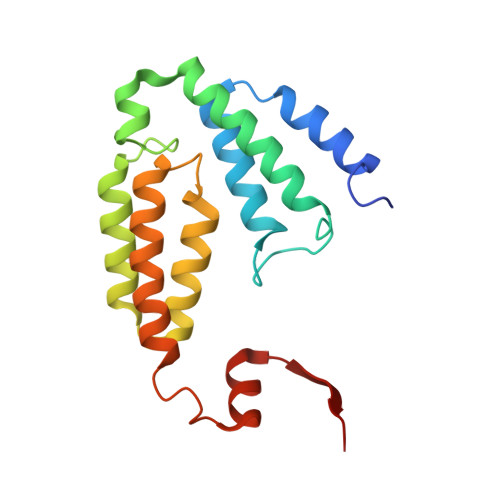
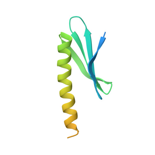
MMOD-induced structural changes of hydroxylase in soluble methane monooxygenase.

Kim, H.An, S.Park, Y.R.Jang, H.Yoo, H.Park, S.H.Lee, S.J.Cho, U.S.

(2019) Sci Adv 5: eaax0059-eaax0059

  • DOI: https://doi.org/10.1126/sciadv.aax0059
  • Primary Citation of Related Structures:  
    6D7K

  • PubMed Abstract: 

    Soluble methane monooxygenase in methanotrophs converts methane to methanol under ambient conditions. The maximum catalytic activity of hydroxylase (MMOH) is achieved through the interplay of its regulatory protein (MMOB) and reductase. An additional auxiliary protein, MMOD, functions as an inhibitor of MMOH; however, its inhibitory mechanism remains unknown. Here, we report the crystal structure of the MMOH-MMOD complex from Methylosinus sporium strain 5 (2.6 Å). Its structure illustrates that MMOD associates with the canyon region of MMOH where MMOB binds. Although MMOD and MMOB recognize the same binding site, each binding component triggers different conformational changes toward MMOH, which then respectively lead to the inhibition and activation of MMOH. Particularly, MMOD binding perturbs the di-iron geometry by inducing two major MMOH conformational changes, i.e., MMOH β subunit disorganization and subsequent His 147 dissociation with Fe1 coordination. Furthermore, 1,6-hexanediol, a mimic of the products of sMMO, reveals the substrate access route.


  • Organizational Affiliation

    Department of Biological Chemistry, University of Michigan, Ann Arbor, MI 48109, USA.


Macromolecules
Find similar proteins by:  (by identity cutoff)  |  3D Structure
Entity ID: 1
MoleculeChains Sequence LengthOrganismDetailsImage
Methane monooxygenase hydroxylase, MmoX1
A, E
526Methylosinus sporiumMutation(s): 0 
UniProt
Find proteins for Q27RN7 (Methylosinus sporium)
Explore Q27RN7 
Go to UniProtKB:  Q27RN7
Entity Groups  
Sequence Clusters30% Identity50% Identity70% Identity90% Identity95% Identity100% Identity
UniProt GroupQ27RN7
Sequence Annotations
Expand
  • Reference Sequence
Find similar proteins by:  (by identity cutoff)  |  3D Structure
Entity ID: 2
MoleculeChains Sequence LengthOrganismDetailsImage
Methane monooxygenase hydroxylase, MmoY
B, F
395Methylosinus sporiumMutation(s): 0 
UniProt
Find proteins for Q27RN6 (Methylosinus sporium)
Explore Q27RN6 
Go to UniProtKB:  Q27RN6
Entity Groups  
Sequence Clusters30% Identity50% Identity70% Identity90% Identity95% Identity100% Identity
UniProt GroupQ27RN6
Sequence Annotations
Expand
  • Reference Sequence
Find similar proteins by:  (by identity cutoff)  |  3D Structure
Entity ID: 3
MoleculeChains Sequence LengthOrganismDetailsImage
Methane monooxygenase hydroxylase, MmoZ
C, G
169Methylosinus sporiumMutation(s): 0 
UniProt
Find proteins for Q27RN4 (Methylosinus sporium)
Explore Q27RN4 
Go to UniProtKB:  Q27RN4
Entity Groups  
Sequence Clusters30% Identity50% Identity70% Identity90% Identity95% Identity100% Identity
UniProt GroupQ27RN4
Sequence Annotations
Expand
  • Reference Sequence
Find similar proteins by:  (by identity cutoff)  |  3D Structure
Entity ID: 4
MoleculeChains Sequence LengthOrganismDetailsImage
Methane monooxygenase hydroxylase, MmoD
D, H
114Methylosinus sporiumMutation(s): 0 
Gene Names: mmoD
UniProt
Find proteins for Q27RN3 (Methylosinus sporium)
Explore Q27RN3 
Go to UniProtKB:  Q27RN3
Entity Groups  
Sequence Clusters30% Identity50% Identity70% Identity90% Identity95% Identity100% Identity
UniProt GroupQ27RN3
Sequence Annotations
Expand
  • Reference Sequence
Small Molecules
Experimental Data & Validation

Experimental Data

  • Method: X-RAY DIFFRACTION
  • Resolution: 2.60 Å
  • R-Value Free: 0.221 
  • R-Value Work: 0.178 
  • R-Value Observed: 0.180 
  • Space Group: C 1 2 1
Unit Cell:
Length ( Å )Angle ( ˚ )
a = 179.781α = 90
b = 125.776β = 102.89
c = 126.38γ = 90
Software Package:
Software NamePurpose
PHENIXrefinement
HKL-2000data reduction
HKL-2000data scaling
PHASERphasing
SCALEPACKdata scaling

Structure Validation

View Full Validation Report


Entry History & Funding Information

Deposition Data


Funding OrganizationLocationGrant Number
National Institutes of Health/National Center for Research Resources (NIH/NCRR)United StatesR01 DK111465
National Research Foundation (NRF, Korea)Korea, Republic OfNRF-2015M3D3A1A01064876

Revision History  (Full details and data files)

  • Version 1.0: 2019-06-26
    Type: Initial release
  • Version 1.1: 2019-10-16
    Changes: Data collection, Database references
  • Version 1.2: 2019-10-30
    Changes: Data collection, Database references
  • Version 1.3: 2019-12-04
    Changes: Author supporting evidence
  • Version 1.4: 2023-10-04
    Changes: Data collection, Database references, Derived calculations, Refinement description