6CG0

Cryo-EM structure of mouse RAG1/2 HFC complex (3.17 A)


Experimental Data Snapshot

  • Method: ELECTRON MICROSCOPY
  • Resolution: 3.17 Å
  • Aggregation State: PARTICLE 
  • Reconstruction Method: SINGLE PARTICLE 

wwPDB Validation   3D Report Full Report


This is version 1.3 of the entry. See complete history


Literature

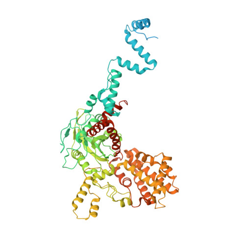
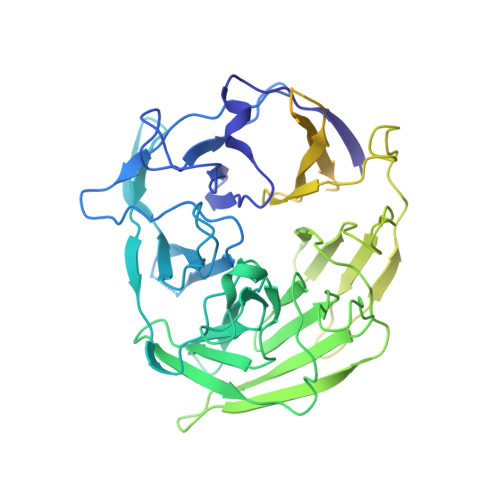
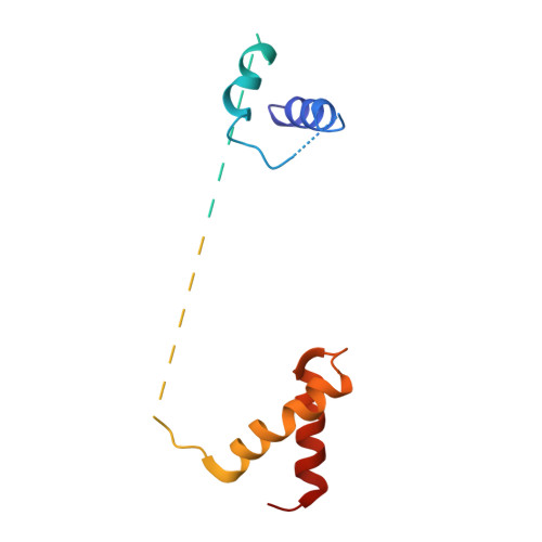
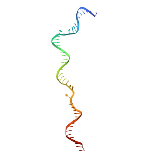
Cracking the DNA Code for V(D)J Recombination.

Kim, M.S.Chuenchor, W.Chen, X.Cui, Y.Zhang, X.Zhou, Z.H.Gellert, M.Yang, W.

(2018) Mol Cell 70: 358-370.e4

  • DOI: 10.1016/j.molcel.2018.03.008
  • Primary Citation of Related Structures:  
    5ZDZ, 5ZE0, 5ZE1, 5ZE2, 6CG0, 6CIJ, 6CIK, 6CIL, 6CIM

  • PubMed Abstract: 
  • To initiate V(D)J recombination for generating the adaptive immune response of vertebrates, RAG1/2 recombinase cleaves DNA at a pair of recombination signal sequences, the 12- and 23-RSS. We have determined crystal and cryo-EM structures of RAG1/2 with DNA in the pre-reaction and hairpin-forming complexes up to 2 ...

    To initiate V(D)J recombination for generating the adaptive immune response of vertebrates, RAG1/2 recombinase cleaves DNA at a pair of recombination signal sequences, the 12- and 23-RSS. We have determined crystal and cryo-EM structures of RAG1/2 with DNA in the pre-reaction and hairpin-forming complexes up to 2.75 Å resolution. Both protein and DNA exhibit structural plasticity and undergo dramatic conformational changes. Coding-flank DNAs extensively rotate, shift, and deform for nicking and hairpin formation. Two intertwined RAG1 subunits crisscross four times between the asymmetric pair of severely bent 12/23-RSS DNAs. Location-sensitive bending of 60° and 150° in 12- and 23-RSS spacers, respectively, must occur for RAG1/2 to capture the nonamers and pair the heptamers for symmetric double-strand breakage. DNA pairing is thus sequence-context dependent and structure specific, which partly explains the "beyond 12/23" restriction. Finally, catalysis in crystallo reveals the process of DNA hairpin formation and its stabilization by interleaved base stacking.


    Organizational Affiliation

    Laboratory of Molecular Biology, NIDDK, NIH, Bethesda, MD 20892, USA. Electronic address: weiy@niddk.nih.gov.


Macromolecules

Find similar proteins by:  (by identity cutoff)  |  3D Structure
Entity ID: 1
MoleculeChainsSequence LengthOrganismDetailsImage
V(D)J recombination-activating protein 1A,
B [auth C]
775Mus musculusMutation(s): 0 
Gene Names: Rag1
EC: 3.1 (PDB Primary Data), 2.3.2.27 (PDB Primary Data)
UniProt
Find proteins for P15919 (Mus musculus)
Explore P15919 
Go to UniProtKB:  P15919
Entity Groups  
Sequence Clusters30% Identity50% Identity70% Identity90% Identity95% Identity100% Identity
UniProt GroupP15919
Protein Feature View
Expand
  • Reference Sequence
Find similar proteins by:  (by identity cutoff)  |  3D Structure
Entity ID: 2
MoleculeChainsSequence LengthOrganismDetailsImage
V(D)J recombination-activating protein 2D [auth B],
C [auth D]
520Mus musculusMutation(s): 0 
Gene Names: Rag2Rag-2
UniProt
Find proteins for P21784 (Mus musculus)
Explore P21784 
Go to UniProtKB:  P21784
Entity Groups  
Sequence Clusters30% Identity50% Identity70% Identity90% Identity95% Identity100% Identity
UniProt GroupP21784
Protein Feature View
Expand
  • Reference Sequence
Find similar proteins by:  (by identity cutoff)  |  3D Structure
Entity ID: 9
MoleculeChainsSequence LengthOrganismDetailsImage
High mobility group protein B1K [auth N]126Homo sapiensMutation(s): 0 
Gene Names: HMGB1HMG1
UniProt & NIH Common Fund Data Resources
Find proteins for P09429 (Homo sapiens)
Explore P09429 
Go to UniProtKB:  P09429
PHAROS:  P09429
Entity Groups  
Sequence Clusters30% Identity50% Identity70% Identity90% Identity95% Identity100% Identity
UniProt GroupP09429
Protein Feature View
Expand
  • Reference Sequence
Find similar nucleic acids by:  (by identity cutoff)  |  3D Structure
Entity ID: 3
MoleculeChainsLengthOrganismImage
DNA (46-MER)E [auth F]46Escherichia coli K-12
Protein Feature View
Expand
  • Reference Sequence

Find similar nucleic acids by:  Sequence   |   3D Structure  

Entity ID: 4
MoleculeChainsLengthOrganismImage
DNA (5'-D(*GP*AP*TP*CP*TP*GP*GP*CP*CP*TP*GP*TP*CP*TP*TP*A)-3')F [auth I]16Homo sapiens
Protein Feature View
Expand
  • Reference Sequence

Find similar nucleic acids by:  Sequence   |   3D Structure  

Entity ID: 5
MoleculeChainsLengthOrganismImage
DNA (5'-D(P*CP*TP*GP*GP*AP*TP*CP*TP*GP*GP*CP*CP*TP*GP*TP*CP*TP*TP*A)-3')G [auth J]19Escherichia coli K-12
Protein Feature View
Expand
  • Reference Sequence
Find similar nucleic acids by:  (by identity cutoff)  |  3D Structure
Entity ID: 6
MoleculeChainsLengthOrganismImage
DNA (60-MER)H [auth G]60Escherichia coli K-12
Protein Feature View
Expand
  • Reference Sequence
Find similar nucleic acids by:  (by identity cutoff)  |  3D Structure
Entity ID: 7
MoleculeChainsLengthOrganismImage
DNA (30-MER)I [auth L]30Escherichia coli K-12
Protein Feature View
Expand
  • Reference Sequence
Find similar nucleic acids by:  (by identity cutoff)  |  3D Structure
Entity ID: 8
MoleculeChainsLengthOrganismImage
DNA (41-MER)J [auth M]41Escherichia coli K-12
Protein Feature View
Expand
  • Reference Sequence
Small Molecules
Ligands 2 Unique
IDChainsName / Formula / InChI Key2D Diagram3D Interactions
ZN
Query on ZN

Download Ideal Coordinates CCD File 
L [auth A],
N [auth C]
ZINC ION
Zn
PTFCDOFLOPIGGS-UHFFFAOYSA-N
 Ligand Interaction
CA
Query on CA

Download Ideal Coordinates CCD File 
M [auth A],
O [auth C]
CALCIUM ION
Ca
BHPQYMZQTOCNFJ-UHFFFAOYSA-N
 Ligand Interaction
Experimental Data & Validation

Experimental Data

  • Method: ELECTRON MICROSCOPY
  • Resolution: 3.17 Å
  • Aggregation State: PARTICLE 
  • Reconstruction Method: SINGLE PARTICLE 
EM Software:
TaskSoftware PackageVersion
RECONSTRUCTIONRELION2.1

Structure Validation

View Full Validation Report


Entry History & Funding Information

Deposition Data


Funding OrganizationLocationGrant Number
National Institutes of Health/National Institute of Diabetes and Digestive and Kidney Disease (NIH/NIDDK)United States1ZIADK036167-11

Revision History  (Full details and data files)

  • Version 1.0: 2018-04-25
    Type: Initial release
  • Version 1.1: 2018-05-02
    Changes: Data collection, Database references
  • Version 1.2: 2019-02-20
    Changes: Author supporting evidence, Data collection
  • Version 1.3: 2019-12-18
    Changes: Author supporting evidence, Other