6BCY

Complex of 14-3-3 theta with an IRSp53 peptide phosphorylated at T360


Experimental Data Snapshot

  • Method: X-RAY DIFFRACTION
  • Resolution: 2.30 Å
  • R-Value Free: 0.222 
  • R-Value Work: 0.192 
  • R-Value Observed: 0.193 

wwPDB Validation   3D Report Full Report


This is version 1.3 of the entry. See complete history


Literature

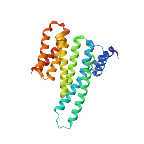
Mechanism of IRSp53 inhibition by 14-3-3.

Kast, D.J.Dominguez, R.

(2019) Nat Commun 10: 483-483

  • DOI: https://doi.org/10.1038/s41467-019-08317-8
  • Primary Citation of Related Structures:  
    6BCR, 6BCY, 6BD1, 6BD2, 6BQT

  • PubMed Abstract: 

    Filopodia are precursors of dendritic spines and polarized cell migration. The I-BAR-domain protein IRSp53 is a key regulator of filopodia dynamics that couples Rho-GTPase signaling to cytoskeleton and membrane remodeling, playing essential roles in neuronal development and cell motility. Here, we describe the structural-functional basis for 14-3-3-dependent inhibition of IRSp53. Phosphoproteomics, quantitative binding and crystallographic studies demonstrate that 14-3-3 binds to two pairs of phosphorylation sites in IRSp53. Using bicistronic expression, we obtain an IRSp53 heterodimer in which only one subunit is phosphorylated, and show that each subunit of IRSp53 independently binds one 14-3-3 dimer. A FRET-sensor assay using natively phosphorylated IRSp53 reveals opposite conformational changes upon binding of activatory (Cdc42, Eps8) or inhibitory (14-3-3) inputs. Finally, we show that 14-3-3 inhibits IRSp53 binding to membranes. Collectively, our findings support a mechanism whereby phosphorylation-dependent inhibition of IRSp53 by 14-3-3 counters membrane binding and interactions with Cdc42 and downstream cytoskeletal effectors.


  • Organizational Affiliation

    Department of Physiology, Perelman School of Medicine, University of Pennsylvania, Philadelphia, PA, 19104, USA.


Macromolecules
Find similar proteins by:  (by identity cutoff)  |  3D Structure
Entity ID: 1
MoleculeChains Sequence LengthOrganismDetailsImage
14-3-3 protein theta
A, B, E, F
245Homo sapiensMutation(s): 0 
UniProt & NIH Common Fund Data Resources
Find proteins for P27348 (Homo sapiens)
Explore P27348 
Go to UniProtKB:  P27348
PHAROS:  P27348
GTEx:  ENSG00000134308 
Entity Groups  
Sequence Clusters30% Identity50% Identity70% Identity90% Identity95% Identity100% Identity
UniProt GroupP27348
Sequence Annotations
Expand
  • Reference Sequence

Find similar proteins by:  Sequence   |   3D Structure  

Entity ID: 2
MoleculeChains Sequence LengthOrganismDetailsImage
Insulin receptor substrate protein of 53 kDa, peptide (IRSp53)
C, D, G, H
13Homo sapiensMutation(s): 0 
UniProt & NIH Common Fund Data Resources
Find proteins for Q9UQB8 (Homo sapiens)
Explore Q9UQB8 
Go to UniProtKB:  Q9UQB8
PHAROS:  Q9UQB8
GTEx:  ENSG00000175866 
Entity Groups  
Sequence Clusters30% Identity50% Identity70% Identity90% Identity95% Identity100% Identity
UniProt GroupQ9UQB8
Sequence Annotations
Expand
  • Reference Sequence
Small Molecules
Ligands 5 Unique
IDChains Name / Formula / InChI Key2D Diagram3D Interactions
PE4
Query on PE4

Download Ideal Coordinates CCD File 
O [auth E]2-{2-[2-(2-{2-[2-(2-ETHOXY-ETHOXY)-ETHOXY]-ETHOXY}-ETHOXY)-ETHOXY]-ETHOXY}-ETHANOL
C16 H34 O8
PJWQOENWHPEPKI-UHFFFAOYSA-N
1PE
Query on 1PE

Download Ideal Coordinates CCD File 
J [auth A],
K [auth B]
PENTAETHYLENE GLYCOL
C10 H22 O6
JLFNLZLINWHATN-UHFFFAOYSA-N
PG4
Query on PG4

Download Ideal Coordinates CCD File 
L [auth B],
P [auth F],
Q [auth F]
TETRAETHYLENE GLYCOL
C8 H18 O5
UWHCKJMYHZGTIT-UHFFFAOYSA-N
PEG
Query on PEG

Download Ideal Coordinates CCD File 
I [auth A],
N [auth E]
DI(HYDROXYETHYL)ETHER
C4 H10 O3
MTHSVFCYNBDYFN-UHFFFAOYSA-N
MG
Query on MG

Download Ideal Coordinates CCD File 
M [auth D],
R [auth H]
MAGNESIUM ION
Mg
JLVVSXFLKOJNIY-UHFFFAOYSA-N
Modified Residues  1 Unique
IDChains TypeFormula2D DiagramParent
TPO
Query on TPO
C, D, G, H
L-PEPTIDE LINKINGC4 H10 N O6 PTHR
Experimental Data & Validation

Experimental Data

  • Method: X-RAY DIFFRACTION
  • Resolution: 2.30 Å
  • R-Value Free: 0.222 
  • R-Value Work: 0.192 
  • R-Value Observed: 0.193 
  • Space Group: P 1
Unit Cell:
Length ( Å )Angle ( ˚ )
a = 59.37α = 72.91
b = 68.63β = 85.59
c = 84.79γ = 64.65
Software Package:
Software NamePurpose
PHENIXrefinement
XDSdata processing
XSCALEdata scaling
PHASERphasing
PDB_EXTRACTdata extraction

Structure Validation

View Full Validation Report


Entry History & Funding Information

Deposition Data


Funding OrganizationLocationGrant Number
National Institutes of Health/National Institute of Mental Health (NIH/NIMH)United StatesR01 MH087950

Revision History  (Full details and data files)

  • Version 1.0: 2018-10-24
    Type: Initial release
  • Version 1.1: 2019-05-08
    Changes: Data collection, Database references
  • Version 1.2: 2019-11-27
    Changes: Author supporting evidence
  • Version 1.3: 2023-10-04
    Changes: Data collection, Database references, Derived calculations, Refinement description