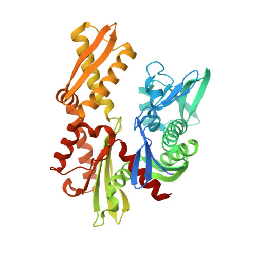
6B1I

Disrupted hydrogen bond network impairs ATPase activity in an Hsc70 cysteine mutant

  • Classification: CHAPERONE
  • Organism(s): Homo sapiens
  • Expression System: Escherichia coli
  • Mutation(s): No 

  • Deposited: 2017-09-18 Released: 2018-01-17 
  • Deposition Author(s): O'Donnell, J.P.
  • Funding Organization(s): National Institutes of Health/National Institute of General Medical Sciences (NIH/NIGMS)

Experimental Data Snapshot

  • Method: X-RAY DIFFRACTION
  • Resolution: 2.30 Å
  • R-Value Free: 0.242 
  • R-Value Work: 0.191 
  • R-Value Observed: 0.194 

wwPDB Validation   3D Report Full Report


Ligand Structure Quality Assessment 


This is version 1.3 of the entry. See complete history


Literature

Disrupted Hydrogen-Bond Network and Impaired ATPase Activity in an Hsc70 Cysteine Mutant.

O'Donnell, J.P.Marsh, H.M.Sondermann, H.Sevier, C.S.

(2018) Biochemistry 57: 1073-1086

  • DOI: https://doi.org/10.1021/acs.biochem.7b01005
  • Primary Citation of Related Structures:  
    6B1I, 6B1M, 6B1N

  • PubMed Abstract: 

    The ATPase domain of members of the 70 kDa heat shock protein (Hsp70) family shows a high degree of sequence, structural, and functional homology across species. A broadly conserved residue within the Hsp70 ATPase domain that captured our attention is an unpaired cysteine, positioned proximal to the site of nucleotide binding. Prior studies of several Hsp70 family members show this cysteine is not required for Hsp70 ATPase activity, yet select amino acid replacements of the cysteine can dramatically alter ATP hydrolysis. Moreover, post-translational modification of the cysteine has been reported to limit ATP hydrolysis for several Hsp70s. To better understand the underlying mechanism for how perturbation of this noncatalytic residue modulates Hsp70 function, we determined the structure for a cysteine-to-tryptophan mutation in the constitutively expressed, mammalian Hsp70 family member Hsc70. Our work reveals that the steric hindrance produced by a cysteine-to-tryptophan mutation disrupts the hydrogen-bond network within the active site, resulting in a loss of proper catalytic magnesium coordination. We propose that a similarly altered active site is likely observed upon post-translational oxidation. We speculate that the subtle changes we detect in the hydrogen-bonding network may relate to the previously reported observation that cysteine oxidation can influence Hsp70 interdomain communication.


  • Organizational Affiliation

    Department of Molecular Medicine, Cornell University , Ithaca, New York 14853, United States.


Macromolecules
Find similar proteins by:  (by identity cutoff)  |  3D Structure
Entity ID: 1
MoleculeChains Sequence LengthOrganismDetailsImage
Heat shock protein family A (Hsp70) member 8
A, B
400Homo sapiensMutation(s): 0 
UniProt & NIH Common Fund Data Resources
Find proteins for P11142 (Homo sapiens)
Explore P11142 
Go to UniProtKB:  P11142
PHAROS:  P11142
GTEx:  ENSG00000109971 
Entity Groups  
Sequence Clusters30% Identity50% Identity70% Identity90% Identity95% Identity100% Identity
UniProt GroupP11142
Sequence Annotations
Expand
  • Reference Sequence
Small Molecules
Ligands 4 Unique
IDChains Name / Formula / InChI Key2D Diagram3D Interactions
ADP (Subject of Investigation/LOI)
Query on ADP

Download Ideal Coordinates CCD File 
E [auth A],
J [auth B]
ADENOSINE-5'-DIPHOSPHATE
C10 H15 N5 O10 P2
XTWYTFMLZFPYCI-KQYNXXCUSA-N
PO4 (Subject of Investigation/LOI)
Query on PO4

Download Ideal Coordinates CCD File 
D [auth A],
I [auth B]
PHOSPHATE ION
O4 P
NBIIXXVUZAFLBC-UHFFFAOYSA-K
GOL
Query on GOL

Download Ideal Coordinates CCD File 
F [auth A],
G [auth A],
K [auth B]
GLYCEROL
C3 H8 O3
PEDCQBHIVMGVHV-UHFFFAOYSA-N
MG (Subject of Investigation/LOI)
Query on MG

Download Ideal Coordinates CCD File 
C [auth A],
H [auth B]
MAGNESIUM ION
Mg
JLVVSXFLKOJNIY-UHFFFAOYSA-N
Binding Affinity Annotations 
IDSourceBinding Affinity
ADP BindingDB:  6B1I Kd: min: 320, max: 324 (nM) from 2 assay(s)
IC50: 280 (nM) from 1 assay(s)
Experimental Data & Validation

Experimental Data

  • Method: X-RAY DIFFRACTION
  • Resolution: 2.30 Å
  • R-Value Free: 0.242 
  • R-Value Work: 0.191 
  • R-Value Observed: 0.194 
  • Space Group: P 1 21 1
Unit Cell:
Length ( Å )Angle ( ˚ )
a = 73.83α = 90
b = 79.196β = 101.45
c = 76.252γ = 90
Software Package:
Software NamePurpose
PHENIXrefinement
XDSdata reduction
SCALAdata scaling
PHASERphasing

Structure Validation

View Full Validation Report


Ligand Structure Quality Assessment 


Entry History & Funding Information

Deposition Data


Funding OrganizationLocationGrant Number
National Institutes of Health/National Institute of General Medical Sciences (NIH/NIGMS)United StatesGM105958

Revision History  (Full details and data files)

  • Version 1.0: 2018-01-17
    Type: Initial release
  • Version 1.1: 2018-02-28
    Changes: Database references
  • Version 1.2: 2020-01-01
    Changes: Author supporting evidence
  • Version 1.3: 2023-10-04
    Changes: Data collection, Database references, Derived calculations, Refinement description