6ARQ

Crystal structure of CD96 (D1) bound to CD155/necl-5 (D1-3)


Experimental Data Snapshot

  • Method: X-RAY DIFFRACTION
  • Resolution: 2.88 Å
  • R-Value Free: 0.252 
  • R-Value Work: 0.230 
  • R-Value Observed: 0.231 

wwPDB Validation   3D Report Full Report


Ligand Structure Quality Assessment 


This is version 2.1 of the entry. See complete history


Literature

Structural Basis for CD96 Immune Receptor Recognition of Nectin-like Protein-5, CD155.

Deuss, F.A.Watson, G.M.Fu, Z.Rossjohn, J.Berry, R.

(2019) Structure 27: 219-228.e3

  • DOI: https://doi.org/10.1016/j.str.2018.10.023
  • Primary Citation of Related Structures:  
    6ARQ

  • PubMed Abstract: 

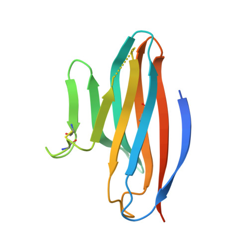
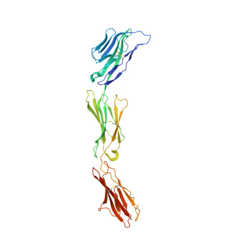
    CD96, DNAM-1, and TIGIT constitute a group of immunoglobulin superfamily receptors that are key regulators of tumor immune surveillance. Within this axis, CD96 recognizes the adhesion molecule nectin-like protein-5 (necl-5), although the molecular basis underpinning this interaction remains unclear. We show that the first immunoglobulin domain (D1) of CD96 is sufficient to mediate a robust interaction with necl-5, but not the DNAM-1 and TIGIT ligand, nectin-2. The crystal structure of CD96-D1 bound to the necl-5 ectodomain revealed that CD96 recognized necl-5 D1 via a conserved "lock-and-key" interaction observed across TIGIT:necl complexes. Specific necl-5 recognition was underpinned by a novel structural motif within CD96, namely an "ancillary key". Mutational analysis showed that this specific residue was critical for necl-5 binding, while simultaneously providing insights into the unique ligand specificity of CD96.


  • Organizational Affiliation

    Infection and Immunity Program and The Department of Biochemistry and Molecular Biology, Biomedicine Discovery Institute, Monash University, Clayton, VIC 3800, Australia; Australian Research Council Centre of Excellence in Advanced Molecular Imaging, Monash University, Clayton, VIC 3800, Australia.


Macromolecules
Find similar proteins by:  (by identity cutoff)  |  3D Structure
Entity ID: 1
MoleculeChains Sequence LengthOrganismDetailsImage
T-cell surface protein tactile128Homo sapiensMutation(s): 1 
Gene Names: CD96
UniProt & NIH Common Fund Data Resources
Find proteins for P40200 (Homo sapiens)
Explore P40200 
Go to UniProtKB:  P40200
PHAROS:  P40200
GTEx:  ENSG00000153283 
Entity Groups  
Sequence Clusters30% Identity50% Identity70% Identity90% Identity95% Identity100% Identity
UniProt GroupP40200
Sequence Annotations
Expand
  • Reference Sequence
Find similar proteins by:  (by identity cutoff)  |  3D Structure
Entity ID: 2
MoleculeChains Sequence LengthOrganismDetailsImage
Poliovirus receptorB [auth D]317Homo sapiensMutation(s): 0 
Gene Names: PVRPVS
UniProt & NIH Common Fund Data Resources
Find proteins for P15151 (Homo sapiens)
Explore P15151 
Go to UniProtKB:  P15151
PHAROS:  P15151
GTEx:  ENSG00000073008 
Entity Groups  
Sequence Clusters30% Identity50% Identity70% Identity90% Identity95% Identity100% Identity
UniProt GroupP15151
Sequence Annotations
Expand
  • Reference Sequence
Oligosaccharides

Help

Entity ID: 3
MoleculeChains Length2D Diagram Glycosylation3D Interactions
alpha-L-fucopyranose-(1-6)-2-acetamido-2-deoxy-beta-D-glucopyranoseC [auth B],
D [auth C],
E
2N-Glycosylation
Glycosylation Resources
GlyTouCan:  G86851RC
GlyCosmos:  G86851RC
GlyGen:  G86851RC
Experimental Data & Validation

Experimental Data

  • Method: X-RAY DIFFRACTION
  • Resolution: 2.88 Å
  • R-Value Free: 0.252 
  • R-Value Work: 0.230 
  • R-Value Observed: 0.231 
  • Space Group: P 21 21 21
Unit Cell:
Length ( Å )Angle ( ˚ )
a = 33.138α = 90
b = 70.819β = 90
c = 224.928γ = 90
Software Package:
Software NamePurpose
BUSTERrefinement
iMOSFLMdata reduction
SCALAdata scaling
PHASERphasing

Structure Validation

View Full Validation Report


Ligand Structure Quality Assessment 


Entry History & Funding Information

Deposition Data


Funding OrganizationLocationGrant Number
National Health and Medical Research Council (NHMRC, Australia)AustraliaAPP1109901

Revision History  (Full details and data files)

  • Version 1.0: 2018-11-21
    Type: Initial release
  • Version 1.1: 2018-11-28
    Changes: Data collection, Database references
  • Version 1.2: 2018-12-26
    Changes: Data collection, Database references
  • Version 1.3: 2019-02-13
    Changes: Data collection, Database references
  • Version 1.4: 2020-01-08
    Changes: Author supporting evidence, Data collection
  • Version 2.0: 2020-07-29
    Type: Remediation
    Reason: Carbohydrate remediation
    Changes: Atomic model, Data collection, Derived calculations, Structure summary
  • Version 2.1: 2023-10-04
    Changes: Data collection, Database references, Derived calculations, Refinement description, Structure summary