5YY9 | pdb_00005yy9

Crystal structure of Tandem Tudor Domain of human UHRF1 in complex with LIG1-K126me3


Experimental Data Snapshot

  • Method: X-RAY DIFFRACTION
  • Resolution: 2.65 Å
  • R-Value Free: 
    0.288 (Depositor), 0.287 (DCC) 
  • R-Value Work: 
    0.231 (Depositor), 0.233 (DCC) 
  • R-Value Observed: 
    0.237 (Depositor) 

Starting Model: experimental
View more details

wwPDB Validation 3D Report Full Report

Validation slider image for 5YY9

This is version 1.3 of the entry. See complete history

Literature

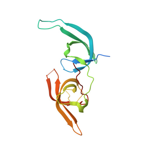
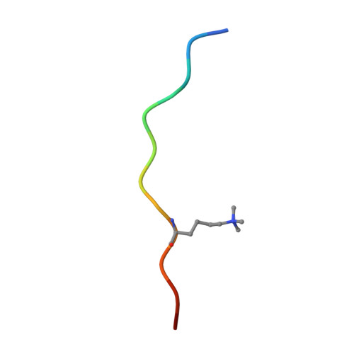
Structure of the UHRF1 Tandem Tudor Domain Bound to a Methylated Non-histone Protein, LIG1, Reveals Rules for Binding and Regulation.

Kori, S.Ferry, L.Matano, S.Jimenji, T.Kodera, N.Tsusaka, T.Matsumura, R.Oda, T.Sato, M.Dohmae, N.Ando, T.Shinkai, Y.Defossez, P.A.Arita, K.

(2019) Structure 27: 485

  • DOI: https://doi.org/10.1016/j.str.2018.11.012
  • Primary Citation Related Structures: 
    5YY9, 5YYA

  • PubMed Abstract: 

    The protein UHRF1 is crucial for DNA methylation maintenance. The tandem Tudor domain (TTD) of UHRF1 binds histone H3K9me2/3 with micromolar affinity, as well as unmethylated linker regions within UHRF1 itself, causing auto-inhibition. Recently, we showed that a methylated histone-like region of DNA ligase 1 (LIG1K126me2/me3) binds the UHRF1 TTD with nanomolar affinity, permitting UHRF1 recruitment to chromatin. Here we report the crystal structure of the UHRF1 TTD bound to a LIG1K126me3 peptide. The data explain the basis for the high TTD-binding affinity of LIG1K126me3 and reveal that the interaction may be regulated by phosphorylation. Binding of LIG1K126me3 switches the overall structure of UHRF1 from a closed to a flexible conformation, suggesting that auto-inhibition is relieved. Our results provide structural insight into how UHRF1 performs its key function in epigenetic maintenance.


  • Organizational Affiliation
    • Graduate School of Medical Life Science, Yokohama City University, Yokohama 230-0045, Japan.

Macromolecule Content 

  • Total Structure Weight: 39.6 kDa 
  • Atom Count: 2,414 
  • Modeled Residue Count: 297 
  • Deposited Residue Count: 334 
  • Unique protein chains: 2

Macromolecules

Find similar proteins by:|  3D Structure
Entity ID: 1
MoleculeChains  Sequence LengthOrganismDetailsImage
E3 ubiquitin-protein ligase UHRF1
A, B
154Homo sapiensMutation(s): 0 
Gene Names: UHRF1ICBP90NP95RNF106
EC: 2.3.2.27
UniProt & NIH Common Fund Data Resources
Find proteins for Q96T88 (Homo sapiens)
Explore Q96T88 
Go to UniProtKB:  Q96T88
PHAROS:  Q96T88
GTEx:  ENSG00000276043 
Entity Groups
Sequence Clusters30% Identity50% Identity70% Identity90% Identity95% Identity100% Identity
UniProt GroupQ96T88
Sequence Annotations
Expand
Reference Sequence
Find similar proteins by:  Sequence   |   3D Structure  
Entity ID: 2
MoleculeChains  Sequence LengthOrganismDetailsImage
Ligase 1
C, D
13synthetic constructMutation(s): 0 
EC: 6.5.1.1
UniProt & NIH Common Fund Data Resources
Find proteins for P18858 (Homo sapiens)
Explore P18858 
Go to UniProtKB:  P18858
PHAROS:  P18858
GTEx:  ENSG00000105486 
Entity Groups
Sequence Clusters30% Identity50% Identity70% Identity90% Identity95% Identity100% Identity
UniProt GroupP18858
Sequence Annotations
Expand
Reference Sequence

Small Molecules

Modified Residues  1 Unique
IDChains TypeFormula2D DiagramParent
M3L
Query on M3L
C, D
L-PEPTIDE LINKINGC9 H21 N2 O2LYS

Experimental Data & Validation

Experimental Data

  • Method: X-RAY DIFFRACTION
  • Resolution: 2.65 Å
  • R-Value Free:  0.288 (Depositor), 0.287 (DCC) 
  • R-Value Work:  0.231 (Depositor), 0.233 (DCC) 
  • R-Value Observed: 0.237 (Depositor) 
Space Group: P 21 21 21
Unit Cell:
Length ( Å )Angle ( ˚ )
a = 27.105α = 90
b = 97.349β = 90
c = 132.529γ = 90
Software Package:
Software NamePurpose
PHENIXrefinement
XDSdata reduction
Aimlessdata scaling
PHASERphasing

Structure Validation

View Full Validation Report


Entry History 

& Funding Information

Deposition Data


Funding OrganizationLocationGrant Number
JSTJapanPRESTO 14530337

Revision History  (Full details and data files)

  • Version 1.0: 2018-12-12
    Type: Initial release
  • Version 1.1: 2019-01-23
    Changes: Data collection, Database references
  • Version 1.2: 2019-03-20
    Changes: Data collection, Database references
  • Version 1.3: 2023-11-22
    Changes: Data collection, Database references, Refinement description