5UMW

Crystal structure of TnmS2, an antibiotic binding protein from Streptomyces sp. CB03234


Experimental Data Snapshot

  • Method: X-RAY DIFFRACTION
  • Resolution: 2.27 Å
  • R-Value Free: 0.263 
  • R-Value Work: 0.205 
  • R-Value Observed: 0.208 

wwPDB Validation   3D Report Full Report


Ligand Structure Quality Assessment 


This is version 1.3 of the entry. See complete history


Literature

Resistance to Enediyne Antitumor Antibiotics by Sequestration.

Chang, C.Y.Yan, X.Crnovcic, I.Annaval, T.Chang, C.Nocek, B.Rudolf, J.D.Yang, D.Babnigg, G.Joachimiak, A.Phillips Jr., G.N.Shen, B.

(2018) Cell Chem Biol 25: 1075-1085.e4

  • DOI: https://doi.org/10.1016/j.chembiol.2018.05.012
  • Primary Citation of Related Structures:  
    5UMP, 5UMQ, 5UMW, 5UMX, 5UMY, 6BBX

  • PubMed Abstract: 

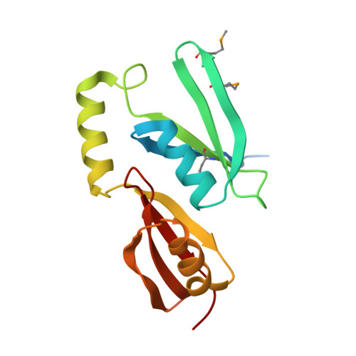
    The enediynes, microbial natural products with extraordinary cytotoxicities, have been translated into clinical drugs. Two self-resistance mechanisms are known in the enediyne producers-apoproteins for the nine-membered enediynes and self-sacrifice proteins for the ten-membered enediyne calicheamicin. Here we show that: (1) tnmS1, tnmS2, and tnmS3 encode tiancimycin (TNM) resistance in its producer Streptomyces sp. CB03234, (2) tnmS1, tnmS2, and tnmS3 homologs are found in all anthraquinone-fused enediyne producers, (3) TnmS1, TnmS2, and TnmS3 share a similar β barrel-like structure, bind TNMs with nanomolar K D values, and confer resistance by sequestration, and (4) TnmS1, TnmS2, and TnmS3 homologs are widespread in nature, including in the human microbiome. These findings unveil an unprecedented resistance mechanism for the enediynes. Mechanisms of self-resistance in producers serve as models to predict and combat future drug resistance in clinical settings. Enediyne-based chemotherapies should now consider the fact that the human microbiome harbors genes encoding enediyne resistance.


  • Organizational Affiliation

    Department of Chemistry, The Scripps Research Institute, Jupiter, FL 33458, USA.


Macromolecules
Find similar proteins by:  (by identity cutoff)  |  3D Structure
Entity ID: 1
MoleculeChains Sequence LengthOrganismDetailsImage
Glyoxalase/bleomycin resisance protein/dioxygenase
A, B, C, D, E
A, B, C, D, E, F
151Streptomyces sp. CB03234Mutation(s): 0 
Gene Names: tnmS2
UniProt
Find proteins for A0A125SA26 (Streptomyces sp. (strain CB03234))
Explore A0A125SA26 
Go to UniProtKB:  A0A125SA26
Entity Groups  
Sequence Clusters30% Identity50% Identity70% Identity90% Identity95% Identity100% Identity
UniProt GroupA0A125SA26
Sequence Annotations
Expand
  • Reference Sequence
Small Molecules
Modified Residues  1 Unique
IDChains TypeFormula2D DiagramParent
MSE
Query on MSE
A, B, C, D, E
A, B, C, D, E, F
L-PEPTIDE LINKINGC5 H11 N O2 SeMET
Binding Affinity Annotations 
IDSourceBinding Affinity
RBF Binding MOAD:  5UMW Kd: 5.95e+4 (nM) from 1 assay(s)
Experimental Data & Validation

Experimental Data

  • Method: X-RAY DIFFRACTION
  • Resolution: 2.27 Å
  • R-Value Free: 0.263 
  • R-Value Work: 0.205 
  • R-Value Observed: 0.208 
  • Space Group: P 32 2 1
Unit Cell:
Length ( Å )Angle ( ˚ )
a = 74.298α = 90
b = 74.298β = 90
c = 342.783γ = 120
Software Package:
Software NamePurpose
REFMACrefinement
AutoSolphasing
HKL-3000data reduction
HKL-3000data scaling

Structure Validation

View Full Validation Report


Ligand Structure Quality Assessment 


Entry History 

Revision History  (Full details and data files)

  • Version 1.0: 2018-07-04
    Type: Initial release
  • Version 1.1: 2018-07-11
    Changes: Data collection, Database references
  • Version 1.2: 2018-10-03
    Changes: Data collection, Database references
  • Version 1.3: 2020-09-23
    Changes: Structure summary