5O7X

CRYSTAL STRUCTURE OF S. CEREVISIAE CORE FACTOR AT 3.2A RESOLUTION


Experimental Data Snapshot

  • Method: X-RAY DIFFRACTION
  • Resolution: 3.20 Å
  • R-Value Free: 0.283 
  • R-Value Work: 0.254 
  • R-Value Observed: 0.255 

wwPDB Validation   3D Report Full Report


This is version 1.2 of the entry. See complete history


Literature

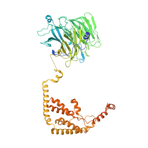
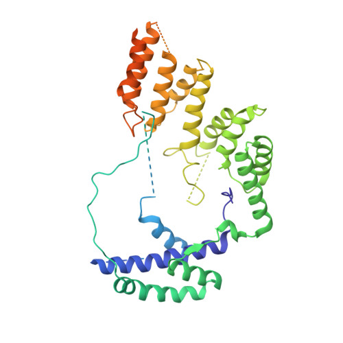
Structural Basis of RNA Polymerase I Transcription Initiation.

Engel, C.Gubbey, T.Neyer, S.Sainsbury, S.Oberthuer, C.Baejen, C.Bernecky, C.Cramer, P.

(2017) Cell 169: 120-131.e22

  • DOI: https://doi.org/10.1016/j.cell.2017.03.003
  • Primary Citation of Related Structures:  
    5N5Y, 5N5Z, 5N60, 5N61, 5O7X

  • PubMed Abstract: 
  • Transcription initiation at the ribosomal RNA promoter requires RNA polymerase (Pol) I and the initiation factors Rrn3 and core factor (CF). Here, we combine X-ray crystallography and cryo-electron microscopy (cryo-EM) to obtain a molecular model for basal Pol I initiation ...

    Transcription initiation at the ribosomal RNA promoter requires RNA polymerase (Pol) I and the initiation factors Rrn3 and core factor (CF). Here, we combine X-ray crystallography and cryo-electron microscopy (cryo-EM) to obtain a molecular model for basal Pol I initiation. The three-subunit CF binds upstream promoter DNA, docks to the Pol I-Rrn3 complex, and loads DNA into the expanded active center cleft of the polymerase. DNA unwinding between the Pol I protrusion and clamp domains enables cleft contraction, resulting in an active Pol I conformation and RNA synthesis. Comparison with the Pol II system suggests that promoter specificity relies on a distinct "bendability" and "meltability" of the promoter sequence that enables contacts between initiation factors, DNA, and polymerase.


    Organizational Affiliation

    Department of Molecular Biology, Max Planck Institute for Biophysical Chemistry, Am Fassberg 11, 37077 Göttingen, Germany. Electronic address: patrick.cramer@mpibpc.mpg.de.


Macromolecules
Find similar proteins by:  (by identity cutoff)  |  3D Structure
Entity ID: 1
MoleculeChainsSequence LengthOrganismDetailsImage
RNA polymerase I-specific transcription initiation factor RRN6
A, D, G, J, M
A, D, G, J, M, P
894Saccharomyces cerevisiae S288CMutation(s): 0 
Gene Names: RRN6YBL014CYBL0311YBL0312
UniProt
Find proteins for P32786 (Saccharomyces cerevisiae (strain ATCC 204508 / S288c))
Explore P32786 
Go to UniProtKB:  P32786
Entity Groups  
Sequence Clusters30% Identity50% Identity70% Identity90% Identity95% Identity100% Identity
UniProt GroupP32786
Protein Feature View
Expand
  • Reference Sequence
Find similar proteins by:  (by identity cutoff)  |  3D Structure
Entity ID: 2
MoleculeChainsSequence LengthOrganismDetailsImage
RNA polymerase I-specific transcription initiation factor RRN7
B, E, H, K, N
B, E, H, K, N, Q
514Saccharomyces cerevisiae S288CMutation(s): 0 
Gene Names: RRN7YJL025WJ1273
UniProt
Find proteins for P40992 (Saccharomyces cerevisiae (strain ATCC 204508 / S288c))
Explore P40992 
Go to UniProtKB:  P40992
Entity Groups  
Sequence Clusters30% Identity50% Identity70% Identity90% Identity95% Identity100% Identity
UniProt GroupP40992
Protein Feature View
Expand
  • Reference Sequence
Find similar proteins by:  (by identity cutoff)  |  3D Structure
Entity ID: 3
MoleculeChainsSequence LengthOrganismDetailsImage
RNA polymerase I-specific transcription initiation factor RRN11
C, F, I, L, O
C, F, I, L, O, R
507Saccharomyces cerevisiae S288CMutation(s): 0 
Gene Names: RRN11YML043CYM9827.09C
UniProt
Find proteins for Q04712 (Saccharomyces cerevisiae (strain ATCC 204508 / S288c))
Explore Q04712 
Go to UniProtKB:  Q04712
Entity Groups  
Sequence Clusters30% Identity50% Identity70% Identity90% Identity95% Identity100% Identity
UniProt GroupQ04712
Protein Feature View
Expand
  • Reference Sequence
Small Molecules
Ligands 2 Unique
IDChainsName / Formula / InChI Key2D Diagram3D Interactions
SO4
Query on SO4

Download Ideal Coordinates CCD File 
AA [auth D]
AB [auth P]
BA [auth D]
DA [auth F]
EA [auth G]
AA [auth D],
AB [auth P],
BA [auth D],
DA [auth F],
EA [auth G],
FA [auth G],
GA [auth G],
IA [auth H],
JA [auth I],
KA [auth J],
LA [auth J],
MA [auth J],
OA [auth K],
PA [auth L],
QA [auth M],
RA [auth M],
S [auth A],
SA [auth M],
T [auth A],
TA [auth M],
U [auth A],
V [auth A],
VA [auth O],
W [auth A],
WA [auth P],
XA [auth P],
Y [auth D],
YA [auth P],
Z [auth D],
ZA [auth P]
SULFATE ION
O4 S
QAOWNCQODCNURD-UHFFFAOYSA-L
 Ligand Interaction
MG
Query on MG

Download Ideal Coordinates CCD File 
BB [auth P]
CA [auth D]
HA [auth G]
NA [auth J]
UA [auth M]
BB [auth P],
CA [auth D],
HA [auth G],
NA [auth J],
UA [auth M],
X [auth A]
MAGNESIUM ION
Mg
JLVVSXFLKOJNIY-UHFFFAOYSA-N
 Ligand Interaction
Experimental Data & Validation

Experimental Data

  • Method: X-RAY DIFFRACTION
  • Resolution: 3.20 Å
  • R-Value Free: 0.283 
  • R-Value Work: 0.254 
  • R-Value Observed: 0.255 
  • Space Group: P 1
Unit Cell:
Length ( Å )Angle ( ˚ )
a = 109.07α = 90.02
b = 109.14β = 90.01
c = 385.64γ = 59.98
Software Package:
Software NamePurpose
PHENIXrefinement
XDSdata reduction
XSCALEdata scaling
PHENIXphasing

Structure Validation

View Full Validation Report


Entry History 

Deposition Data

Revision History  (Full details and data files)

  • Version 1.0: 2017-08-02
    Type: Initial release
  • Version 1.1: 2017-08-09
    Changes: Structure summary
  • Version 1.2: 2019-10-16
    Changes: Data collection