5MYX | pdb_00005myx

Structure of Pyroglutamate-Abeta-specific Fab c#24 in complex with human Abeta-pE3-18


Experimental Data Snapshot

  • Method: X-RAY DIFFRACTION
  • Resolution: 1.49 Å
  • R-Value Free: 
    0.213 (Depositor), 0.213 (DCC) 
  • R-Value Work: 
    0.180 (Depositor), 0.181 (DCC) 
  • R-Value Observed: 
    0.182 (Depositor) 

Starting Model: experimental
View more details

wwPDB Validation 3D Report Full Report

Validation slider image for 5MYX

This is version 2.2 of the entry. See complete history

Literature

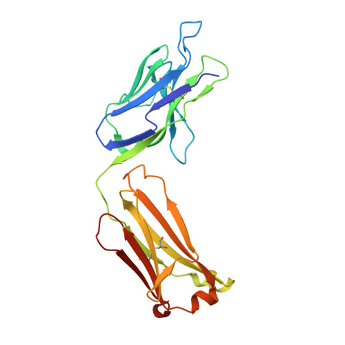
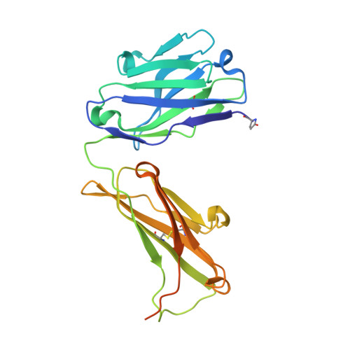
Structural and functional analyses of pyroglutamate-amyloid-beta-specific antibodies as a basis for Alzheimer immunotherapy.

Piechotta, A.Parthier, C.Kleinschmidt, M.Gnoth, K.Pillot, T.Lues, I.Demuth, H.U.Schilling, S.Rahfeld, J.U.Stubbs, M.T.

(2017) J Biological Chem 292: 12713-12724

  • DOI: https://doi.org/10.1074/jbc.M117.777839
  • Primary Citation Related Structures: 
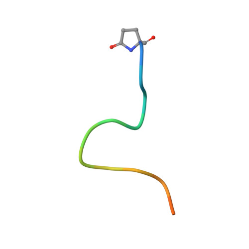
    5MY4, 5MYK, 5MYO, 5MYX

  • PubMed Abstract: 

    Alzheimer disease is associated with deposition of the amyloidogenic peptide Aβ in the brain. Passive immunization using Aβ-specific antibodies has been demonstrated to reduce amyloid deposition both in vitro and in vivo Because N-terminally truncated pyroglutamate (pE)-modified Aβ species (Aβ pE3 ) exhibit enhanced aggregation potential and propensity to form toxic oligomers, they represent particularly attractive targets for antibody therapy. Here we present three separate monoclonal antibodies that specifically recognize Aβ pE3 with affinities of 1-10 nm and inhibit Aβ pE3 fibril formation in vitro. In vivo application of one of these resulted in improved memory in Aβ pE3 oligomer-treated mice. Crystal structures of F ab -Aβ pE3 complexes revealed two distinct binding modes for the peptide. Juxtaposition of pyroglutamate pE3 and the F4 side chain (the "pEF head") confers a pronounced bulky hydrophobic nature to the Aβ pE3 N terminus that might explain the enhanced aggregation properties of the modified peptide. The deep burial of the pEF head by two of the antibodies explains their high target specificity and low cross-reactivity, making them promising candidates for the development of clinical antibodies.


  • Organizational Affiliation
    • Probiodrug AG, Weinbergweg 22, 06120 Halle (Saale), Germany; Institute of Biotechnology, Martin Luther University, 06108 Halle-Wittenberg, Germany; Department of Molecular Drug Biochemistry and Therapy, Fraunhofer Institute for Cell Therapy and Immunology, Weinbergweg 22, 06120 Halle, Germany.

Macromolecule Content 

  • Total Structure Weight: 105.33 kDa 
  • Atom Count: 8,484 
  • Modeled Residue Count: 890 
  • Deposited Residue Count: 966 
  • Unique protein chains: 3

Macromolecules

Find similar proteins by:|  3D Structure
Entity ID: 1
MoleculeChains  Sequence LengthOrganismDetailsImage
Fab c#24 light chain
A, C
219Mus musculusMutation(s): 0 
Find similar proteins by:|  3D Structure
Entity ID: 2
MoleculeChains  Sequence LengthOrganismDetailsImage
Fab c#24 heavy chain
B, D
248Mus musculusMutation(s): 0 
Find similar proteins by:  Sequence   |   3D Structure  
Entity ID: 3
MoleculeChains  Sequence LengthOrganismDetailsImage
Pyroglutamate-Abeta pE3-18
E, F
16Homo sapiensMutation(s): 0 
UniProt & NIH Common Fund Data Resources
Find proteins for P05067 (Homo sapiens)
Explore P05067 
Go to UniProtKB:  P05067
PHAROS:  P05067
GTEx:  ENSG00000142192 
Entity Groups
Sequence Clusters30% Identity50% Identity70% Identity90% Identity95% Identity100% Identity
UniProt GroupP05067
Sequence Annotations
Expand
Reference Sequence

Small Molecules

Modified Residues  1 Unique
IDChains TypeFormula2D DiagramParent
PCA
Query on PCA
B, D
L-PEPTIDE LINKINGC5 H7 N O3GLN

Experimental Data & Validation

Experimental Data

  • Method: X-RAY DIFFRACTION
  • Resolution: 1.49 Å
  • R-Value Free:  0.213 (Depositor), 0.213 (DCC) 
  • R-Value Work:  0.180 (Depositor), 0.181 (DCC) 
  • R-Value Observed: 0.182 (Depositor) 
Space Group: C 1 2 1
Unit Cell:
Length ( Å )Angle ( ˚ )
a = 115.611α = 90
b = 95.935β = 101.17
c = 90.371γ = 90
Software Package:
Software NamePurpose
PHASERphasing
PHENIXrefinement
PDB_EXTRACTdata extraction
XDSdata reduction
XDSdata scaling

Structure Validation

View Full Validation Report


Entry History 

Deposition Data

Revision History  (Full details and data files)

  • Version 1.0: 2017-06-28
    Type: Initial release
  • Version 1.1: 2017-08-09
    Changes: Database references
  • Version 2.0: 2020-03-11
    Changes: Atomic model, Data collection, Polymer sequence
  • Version 2.1: 2024-01-17
    Changes: Data collection, Database references, Refinement description
  • Version 2.2: 2024-11-20
    Changes: Structure summary