5BTA

Crystal structure of a topoisomerase II complex


Experimental Data Snapshot

  • Method: X-RAY DIFFRACTION
  • Resolution: 2.55 Å
  • R-Value Free: 0.251 
  • R-Value Work: 0.223 
  • R-Value Observed: 0.225 

wwPDB Validation   3D Report Full Report


Ligand Structure Quality Assessment 


This is version 1.4 of the entry. See complete history


Literature

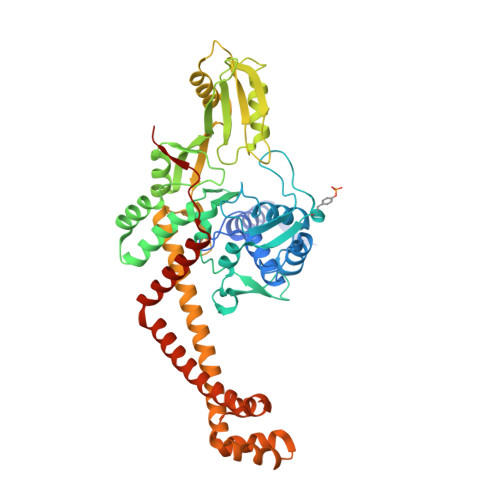
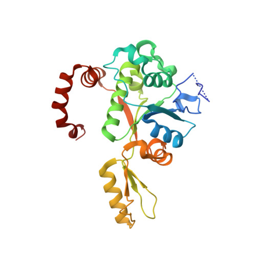
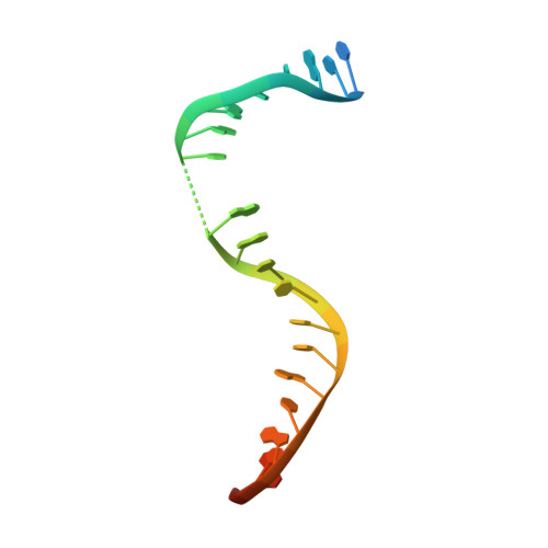
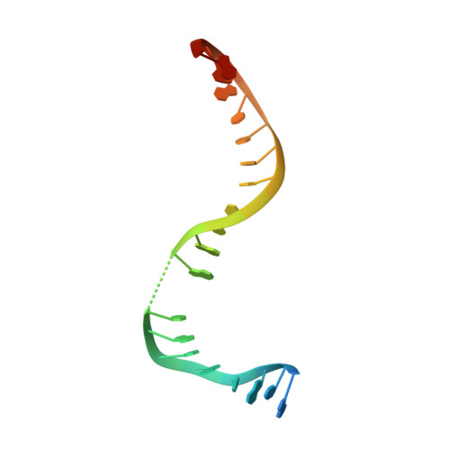
Crystal structure and stability of gyrase-fluoroquinolone cleaved complexes from Mycobacterium tuberculosis.

Blower, T.R.Williamson, B.H.Kerns, R.J.Berger, J.M.

(2016) Proc Natl Acad Sci U S A 113: 1706-1713

  • DOI: https://doi.org/10.1073/pnas.1525047113
  • Primary Citation of Related Structures:  
    5BS8, 5BTA, 5BTC, 5BTD, 5BTF, 5BTG, 5BTI, 5BTL, 5BTN

  • PubMed Abstract: 

    Mycobacterium tuberculosis (Mtb) infects one-third of the world's population and in 2013 accounted for 1.5 million deaths. Fluoroquinolone antibacterials, which target DNA gyrase, are critical agents used to halt the progression from multidrug-resistant tuberculosis to extensively resistant disease; however, fluoroquinolone resistance is emerging and new ways to bypass resistance are required. To better explain known differences in fluoroquinolone action, the crystal structures of the WT Mtb DNA gyrase cleavage core and a fluoroquinolone-sensitized mutant were determined in complex with DNA and five fluoroquinolones. The structures, ranging from 2.4- to 2.6-Å resolution, show that the intrinsically low susceptibility of Mtb to fluoroquinolones correlates with a reduction in contacts to the water shell of an associated magnesium ion, which bridges fluoroquinolone-gyrase interactions. Surprisingly, the structural data revealed few differences in fluoroquinolone-enzyme contacts from drugs that have very different activities against Mtb. By contrast, a stability assay using purified components showed a clear relationship between ternary complex reversibility and inhibitory activities reported with cultured cells. Collectively, our data indicate that the stability of fluoroquinolone/DNA interactions is a major determinant of fluoroquinolone activity and that moieties that have been appended to the C7 position of different quinolone scaffolds do not take advantage of specific contacts that might be made with the enzyme. These concepts point to new approaches for developing quinolone-class compounds that have increased potency against Mtb and the ability to overcome resistance.


  • Organizational Affiliation

    Department of Biophysics and Biophysical Chemistry, Johns Hopkins University School of Medicine, Baltimore, MD 21205;


Macromolecules

Find similar proteins by:  (by identity cutoff)  |  3D Structure
Entity ID: 1
MoleculeChains Sequence LengthOrganismDetailsImage
DNA gyrase subunit A
A, C
503Mycobacterium tuberculosis H37RvMutation(s): 1 
Gene Names: gyrARv0006MTCY10H4.04
EC: 5.99.1.3
UniProt
Find proteins for P9WG47 (Mycobacterium tuberculosis (strain ATCC 25618 / H37Rv))
Explore P9WG47 
Go to UniProtKB:  P9WG47
Entity Groups  
Sequence Clusters30% Identity50% Identity70% Identity90% Identity95% Identity100% Identity
UniProt GroupP9WG47
Sequence Annotations
Expand
  • Reference Sequence
Find similar proteins by:  (by identity cutoff)  |  3D Structure
Entity ID: 2
MoleculeChains Sequence LengthOrganismDetailsImage
DNA gyrase subunit B
B, D
253Mycobacterium tuberculosis H37RvMutation(s): 0 
Gene Names: gyrBRv0005MTCY10H4.03
EC: 5.99.1.3
UniProt
Find proteins for P9WG45 (Mycobacterium tuberculosis (strain ATCC 25618 / H37Rv))
Explore P9WG45 
Go to UniProtKB:  P9WG45
Entity Groups  
Sequence Clusters30% Identity50% Identity70% Identity90% Identity95% Identity100% Identity
UniProt GroupP9WG45
Sequence Annotations
Expand
  • Reference Sequence

Find similar nucleic acids by:  Sequence   |   3D Structure  

Entity ID: 3
MoleculeChains LengthOrganismImage
DNA substrate 24-mer GGTCATGAATGACTATGCACGTAA
E, H
24synthetic construct
Sequence Annotations
Expand
  • Reference Sequence

Find similar nucleic acids by:  Sequence   |   3D Structure  

Entity ID: 4
MoleculeChains LengthOrganismImage
DNA substrate 24-mer TTACGTGCATAGTCATTCATGACC
F, G
24synthetic construct
Sequence Annotations
Expand
  • Reference Sequence
Small Molecules
Modified Residues  1 Unique
IDChains TypeFormula2D DiagramParent
PTR
Query on PTR
A, C
L-PEPTIDE LINKINGC9 H12 N O6 PTYR
Binding Affinity Annotations 
IDSourceBinding Affinity
MFX BindingDB:  5BTA IC50: 5.00e+4 (nM) from 1 assay(s)
Experimental Data & Validation

Experimental Data

  • Method: X-RAY DIFFRACTION
  • Resolution: 2.55 Å
  • R-Value Free: 0.251 
  • R-Value Work: 0.223 
  • R-Value Observed: 0.225 
  • Space Group: P 1 21 1
Unit Cell:
Length ( Å )Angle ( ˚ )
a = 108.34α = 90
b = 83.205β = 108.55
c = 129.757γ = 90
Software Package:
Software NamePurpose
PHENIXrefinement
XSCALEdata scaling
PDB_EXTRACTdata extraction
XDSdata reduction
PHASERphasing
Cootmodel building

Structure Validation

View Full Validation Report


Ligand Structure Quality Assessment 


Entry History & Funding Information

Deposition Data


Funding OrganizationLocationGrant Number
National Institutes of Health/National Cancer Institute (NIH/NCI)United StatesR01-CA077373

Revision History  (Full details and data files)

  • Version 1.0: 2016-03-02
    Type: Initial release
  • Version 1.1: 2017-09-20
    Changes: Author supporting evidence, Database references, Derived calculations
  • Version 1.2: 2019-12-04
    Changes: Author supporting evidence
  • Version 1.3: 2023-09-27
    Changes: Data collection, Database references, Derived calculations, Refinement description
  • Version 1.4: 2023-11-15
    Changes: Data collection