5ZQ6

SidE-Ubi-ADPr


Experimental Data Snapshot

  • Method: X-RAY DIFFRACTION
  • Resolution: 3.01 Å
  • R-Value Free: 0.232 
  • R-Value Work: 0.180 
  • R-Value Observed: 0.183 

wwPDB Validation   3D Report Full Report


This is version 1.2 of the entry. See complete history


Literature

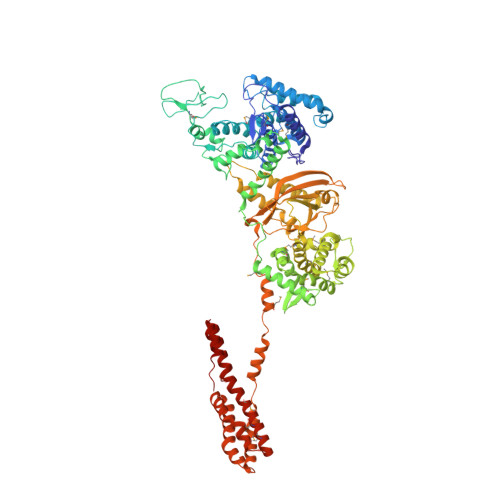
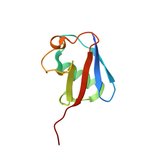
Structural Insights into Non-canonical Ubiquitination Catalyzed by SidE.

Wang, Y.Shi, M.Feng, H.Zhu, Y.Liu, S.Gao, A.Gao, P.

(2018) Cell 173: 1231-1243.e16

  • DOI: https://doi.org/10.1016/j.cell.2018.04.023
  • Primary Citation of Related Structures:  
    5ZQ2, 5ZQ3, 5ZQ4, 5ZQ5, 5ZQ6, 5ZQ7

  • PubMed Abstract: 

    Ubiquitination constitutes one of the most important signaling mechanisms in eukaryotes. Conventional ubiquitination is catalyzed by the universally conserved E1-E2-E3 three-enzyme cascade in an ATP-dependent manner. The newly identified SidE family effectors of the pathogen Legionella pneumophila ubiquitinate several human proteins by a different mechanism without engaging any of the conventional ubiquitination machinery. We now report the crystal structures of SidE alone and in complex with ubiquitin, NAD, and ADP-ribose, thereby capturing different conformations of SidE before and after ubiquitin and ligand binding. The structures of ubiquitin bound to both mART and PDE domains reveal several unique features of the two reaction steps catalyzed by SidE. Further, the structural and biochemical results demonstrate that SidE family members do not recognize specific structural folds of the substrate proteins. Our studies provide both structural explanations for the functional observations and new insights into the molecular mechanisms of this non-canonical ubiquitination machinery.


  • Organizational Affiliation

    CAS Key Laboratory of Infection and Immunity, CAS Center for Excellence in Biomacromolecules, Institute of Biophysics, Chinese Academy of Sciences, Beijing 100101, China.


Macromolecules
Find similar proteins by:  (by identity cutoff)  |  3D Structure
Entity ID: 1
MoleculeChains Sequence LengthOrganismDetailsImage
SidEA,
D [auth C]
845Legionella pneumophilaMutation(s): 0 
UniProt
Find proteins for Q6BBR6 (Legionella pneumophila)
Explore Q6BBR6 
Go to UniProtKB:  Q6BBR6
Entity Groups  
Sequence Clusters30% Identity50% Identity70% Identity90% Identity95% Identity100% Identity
UniProt GroupQ6BBR6
Sequence Annotations
Expand
  • Reference Sequence
Find similar proteins by:  (by identity cutoff)  |  3D Structure
Entity ID: 2
MoleculeChains Sequence LengthOrganismDetailsImage
UbiquitinB,
C [auth D]
79Homo sapiensMutation(s): 1 
Gene Names: UBC
UniProt & NIH Common Fund Data Resources
Find proteins for P0CG48 (Homo sapiens)
Explore P0CG48 
Go to UniProtKB:  P0CG48
PHAROS:  P0CG48
GTEx:  ENSG00000150991 
Entity Groups  
Sequence Clusters30% Identity50% Identity70% Identity90% Identity95% Identity100% Identity
UniProt GroupP0CG48
Sequence Annotations
Expand
  • Reference Sequence
Small Molecules
Modified Residues  1 Unique
IDChains TypeFormula2D DiagramParent
MSE
Query on MSE
A,
D [auth C]
L-PEPTIDE LINKINGC5 H11 N O2 SeMET
Experimental Data & Validation

Experimental Data

  • Method: X-RAY DIFFRACTION
  • Resolution: 3.01 Å
  • R-Value Free: 0.232 
  • R-Value Work: 0.180 
  • R-Value Observed: 0.183 
  • Space Group: P 21 21 21
Unit Cell:
Length ( Å )Angle ( ˚ )
a = 92.322α = 90
b = 128.003β = 90
c = 191.065γ = 90
Software Package:
Software NamePurpose
PHENIXrefinement
Cootmodel building
HKL-3000data scaling
PHENIXphasing

Structure Validation

View Full Validation Report


Entry History & Funding Information

Deposition Data


Funding OrganizationLocationGrant Number
China91753133
China31670903
China31700687
ChinaXDB08020204

Revision History  (Full details and data files)

  • Version 1.0: 2018-05-23
    Type: Initial release
  • Version 1.1: 2018-05-30
    Changes: Data collection, Database references
  • Version 1.2: 2023-11-22
    Changes: Data collection, Database references, Refinement description