5YQS

Isoprimeverose-producing enzyme from Aspergillus oryzae in complex with isoprimeverose


Experimental Data Snapshot

  • Method: X-RAY DIFFRACTION
  • Resolution: 2.40 Å
  • R-Value Free: 0.208 
  • R-Value Work: 0.153 
  • R-Value Observed: 0.156 

wwPDB Validation   3D Report Full Report


Ligand Structure Quality Assessment 


This is version 2.0 of the entry. See complete history


Literature

Crystal structure and substrate recognition mechanism of Aspergillus oryzae isoprimeverose-producing enzyme.

Matsuzawa, T.Watanabe, M.Nakamichi, Y.Fujimoto, Z.Yaoi, K.

(2019) J Struct Biol 205: 84-90

  • DOI: https://doi.org/10.1016/j.jsb.2018.11.005
  • Primary Citation of Related Structures:  
    5YOT, 5YQS

  • PubMed Abstract: 

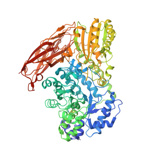
    Isoprimeverose-producing enzymes (IPases) release isoprimeverose (α-d-xylopyranosyl-(1 → 6)-d-glucopyranose) from the non-reducing end of xyloglucan oligosaccharides. Aspergillus oryzae IPase (IpeA) is classified as a member of the glycoside hydrolase family 3 (GH3); however, it has unusual substrate specificity compared with other GH3 enzymes. Xylopyranosyl branching at the non-reducing ends of xyloglucan oligosaccharides is vital for IpeA activity. We solved the crystal structure of IpeA with isoprimeverose at 2.4 Å resolution, showing that the structure of IpeA formed a dimer and was composed of three domains: an N-terminal (β/α) 8 TIM-barrel domain, α/β/α sandwich fold domain, and a C-terminal fibronectin-like domain. The catalytic TIM-barrel domain possessed a catalytic nucleophile (Asp300) and acid/base (Glu524) residues. Interestingly, we found that the cavity of the active site of IpeA was larger than that of other GH3 enzymes, and subsite -1' played an important role in its activity. The glucopyranosyl and xylopyranosyl residues of isoprimeverose were located at subsites -1 and -1', respectively. Gln58 and Tyr89 contributed to the interaction with the xylopyranosyl residue of isoprimeverose through hydrogen bonding and stacking effects, respectively. Our findings provide new insights into the substrate recognition of GH3 enzymes.


  • Organizational Affiliation

    Bioproduction Research Institute, National Institute of Advanced Industrial Science and Technology (AIST), Tsukuba Central 6, 1-1-1 Higashi, Tsukuba, Ibaraki 305-8566, Japan.


Macromolecules
Find similar proteins by:  (by identity cutoff)  |  3D Structure
Entity ID: 1
MoleculeChains Sequence LengthOrganismDetailsImage
Isoprimeverose-producing enzyme
A, B
765Aspergillus oryzae RIB40Mutation(s): 0 
Gene Names: AO090701000274
UniProt
Find proteins for Q2U8V9 (Aspergillus oryzae (strain ATCC 42149 / RIB 40))
Explore Q2U8V9 
Go to UniProtKB:  Q2U8V9
Entity Groups  
Sequence Clusters30% Identity50% Identity70% Identity90% Identity95% Identity100% Identity
UniProt GroupQ2U8V9
Sequence Annotations
Expand
  • Reference Sequence
Oligosaccharides

Help

Entity ID: 2
MoleculeChains Length2D Diagram Glycosylation3D Interactions
2-acetamido-2-deoxy-beta-D-glucopyranose-(1-4)-2-acetamido-2-deoxy-beta-D-glucopyranose
C, D, E, I, J
C, D, E, I, J, K
2N-Glycosylation
Glycosylation Resources
GlyTouCan:  G42666HT
GlyCosmos:  G42666HT
GlyGen:  G42666HT
Entity ID: 3
MoleculeChains Length2D Diagram Glycosylation3D Interactions
alpha-D-xylopyranose-(1-6)-beta-D-glucopyranose
F, H, L, N
2N/A
Glycosylation Resources
GlyTouCan:  G00977CB
GlyCosmos:  G00977CB
GlyGen:  G00977CB
Entity ID: 4
MoleculeChains Length2D Diagram Glycosylation3D Interactions
alpha-D-xylopyranose-(1-6)-alpha-D-glucopyranose
G, M
2N/A
Glycosylation Resources
GlyTouCan:  G00097DN
GlyCosmos:  G00097DN
GlyGen:  G00097DN
Small Molecules
Ligands 3 Unique
IDChains Name / Formula / InChI Key2D Diagram3D Interactions
NAG
Query on NAG

Download Ideal Coordinates CCD File 
O [auth A]
P [auth A]
Q [auth A]
R [auth A]
S [auth A]
O [auth A],
P [auth A],
Q [auth A],
R [auth A],
S [auth A],
V [auth B],
W [auth B],
X [auth B],
Y [auth B]
2-acetamido-2-deoxy-beta-D-glucopyranose
C8 H15 N O6
OVRNDRQMDRJTHS-FMDGEEDCSA-N
BGC
Query on BGC

Download Ideal Coordinates CCD File 
U [auth A]beta-D-glucopyranose
C6 H12 O6
WQZGKKKJIJFFOK-VFUOTHLCSA-N
CA
Query on CA

Download Ideal Coordinates CCD File 
T [auth A],
Z [auth B]
CALCIUM ION
Ca
BHPQYMZQTOCNFJ-UHFFFAOYSA-N
Experimental Data & Validation

Experimental Data

  • Method: X-RAY DIFFRACTION
  • Resolution: 2.40 Å
  • R-Value Free: 0.208 
  • R-Value Work: 0.153 
  • R-Value Observed: 0.156 
  • Space Group: P 1 21 1
Unit Cell:
Length ( Å )Angle ( ˚ )
a = 69.787α = 90
b = 129.911β = 94.44
c = 95.162γ = 90
Software Package:
Software NamePurpose
REFMACrefinement
SCALEPACKdata scaling
PDB_EXTRACTdata extraction
DENZOdata reduction
PHENIXphasing

Structure Validation

View Full Validation Report


Ligand Structure Quality Assessment 


Entry History 

Deposition Data

Revision History  (Full details and data files)

  • Version 1.0: 2018-11-14
    Type: Initial release
  • Version 1.1: 2019-11-13
    Changes: Data collection, Database references
  • Version 2.0: 2020-07-29
    Type: Remediation
    Reason: Carbohydrate remediation
    Changes: Atomic model, Data collection, Derived calculations, Structure summary