5X30

Crystal structure of Pseudomonas putida methionine gamma-lyase C116H mutant with L-homocysteine intermediates.


Experimental Data Snapshot

  • Method: X-RAY DIFFRACTION
  • Resolution: 1.70 Å
  • R-Value Free: 0.195 
  • R-Value Work: 0.170 
  • R-Value Observed: 0.171 

wwPDB Validation   3D Report Full Report


Ligand Structure Quality Assessment 


This is version 1.3 of the entry. See complete history


Literature

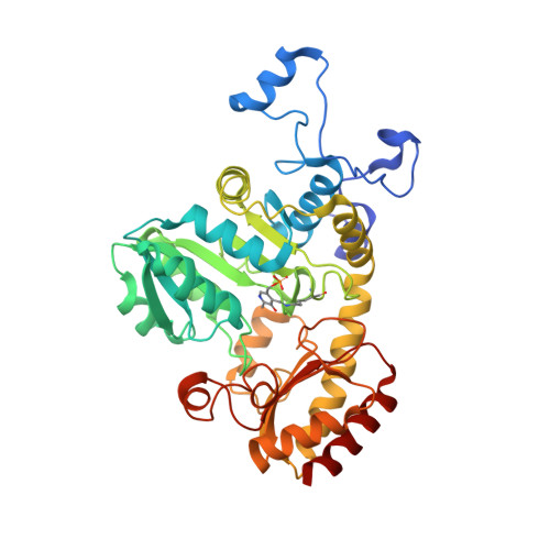
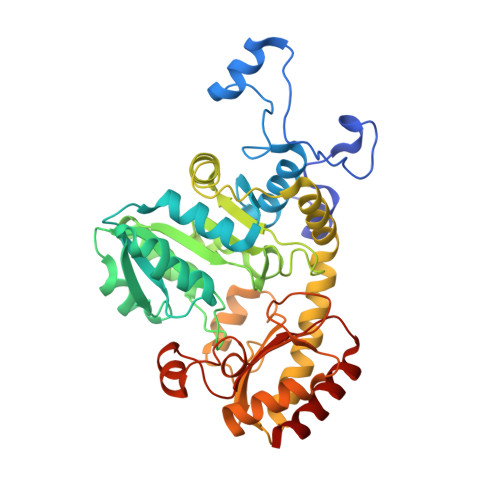
Structural and mechanistic insights into homocysteine degradation by a mutant of methionine gamma-lyase based on substrate-assisted catalysis

Sato, D.Shiba, T.Yunoto, S.Furutani, K.Fukumoto, M.Kudou, D.Tamura, T.Inagaki, K.Harada, S.

(2017) Protein Sci 26: 1224-1230

  • DOI: https://doi.org/10.1002/pro.3158
  • Primary Citation of Related Structures:  
    5X2V, 5X2W, 5X2X, 5X2Y, 5X2Z, 5X30

  • PubMed Abstract: 

    Methionine γ-lyse (MGL) catalyzes the α, γ-elimination of l-methionine and its derivatives as well as the α, β-elimination of l-cysteine and its derivatives to produce α-keto acids, volatile thiols, and ammonia. The reaction mechanism of MGL has been characterized by enzymological studies using several site-directed mutants. The Pseudomonas putida MGL C116H mutant showed drastically reduced degradation activity toward methionine while retaining activity toward homocysteine. To understand the underlying mechanism and to discern the subtle differences between these substrates, we analyzed the crystal structures of the reaction intermediates. The complex formed between the C116H mutant and methionine demonstrated that a loop structure (Ala51-Asn64) in the adjacent subunit of the catalytic dimer cannot approach the cofactor pyridoxal 5'-phosphate (PLP) because His116 disrupts the interaction of Asp241 with Lys240, and the liberated side chain of Lys240 causes steric hindrance with this loop. Conversely, in the complex formed between C116H mutant and homocysteine, the thiol moiety of the substrate conjugated with PLP offsets the imidazole ring of His116 via a water molecule, disrupting the interaction of His116 and Asp241 and restoring the interaction of Asp241 with Lys240. These structural data suggest that the Cys116 to His mutation renders the enzyme inactive toward the original substrate, but activity is restored when the substrate is homocysteine due to substrate-assisted catalysis.


  • Organizational Affiliation

    Department of Applied Biology, Graduate School of Science Technology, Kyoto Institute of Technology, Kyoto, 606-8585, Japan.


Macromolecules
Find similar proteins by:  (by identity cutoff)  |  3D Structure
Entity ID: 1
MoleculeChains Sequence LengthOrganismDetailsImage
L-methionine gamma-lyase
A, C
398Pseudomonas putidaMutation(s): 1 
Gene Names: mdeA
EC: 4.4.1.11 (PDB Primary Data), 4.4.1.2 (PDB Primary Data)
UniProt
Find proteins for P13254 (Pseudomonas putida)
Explore P13254 
Go to UniProtKB:  P13254
Entity Groups  
Sequence Clusters30% Identity50% Identity70% Identity90% Identity95% Identity100% Identity
UniProt GroupP13254
Sequence Annotations
Expand
  • Reference Sequence
Find similar proteins by:  (by identity cutoff)  |  3D Structure
Entity ID: 2
MoleculeChains Sequence LengthOrganismDetailsImage
L-methionine gamma-lyase
B, D
398Pseudomonas putidaMutation(s): 1 
Gene Names: mdeA
EC: 4.4.1.11 (PDB Primary Data), 4.4.1.2 (PDB Primary Data)
UniProt
Find proteins for P13254 (Pseudomonas putida)
Explore P13254 
Go to UniProtKB:  P13254
Entity Groups  
Sequence Clusters30% Identity50% Identity70% Identity90% Identity95% Identity100% Identity
UniProt GroupP13254
Sequence Annotations
Expand
  • Reference Sequence
Small Molecules
Ligands 4 Unique
IDChains Name / Formula / InChI Key2D Diagram3D Interactions
7XF
Query on 7XF

Download Ideal Coordinates CCD File 
I [auth D](2~{S})-2-[[2-methyl-3-oxidanyl-5-(phosphonooxymethyl)pyridin-4-yl]methylamino]-4-sulfanyl-butanoic acid
C12 H19 N2 O7 P S
PXCMMZUTCUCUSM-JTQLQIEISA-N
4LM
Query on 4LM

Download Ideal Coordinates CCD File 
F [auth B](2E)-2-{[(1E)-{3-hydroxy-2-methyl-5-[(phosphonooxy)methyl]pyridin-4-yl}methylidene]amino}but-2-enoic acid
C12 H15 N2 O7 P
BBYSOXSBJOWRNU-VMTXVVAMSA-N
HCS
Query on HCS

Download Ideal Coordinates CCD File 
E [auth A],
H [auth C]
2-AMINO-4-MERCAPTO-BUTYRIC ACID
C4 H9 N O2 S
FFFHZYDWPBMWHY-VKHMYHEASA-N
H2S
Query on H2S

Download Ideal Coordinates CCD File 
G [auth B]HYDROSULFURIC ACID
H2 S
RWSOTUBLDIXVET-UHFFFAOYSA-N
Modified Residues  1 Unique
IDChains TypeFormula2D DiagramParent
LLP
Query on LLP
A, C
L-PEPTIDE LINKINGC14 H22 N3 O7 PLYS
Experimental Data & Validation

Experimental Data

  • Method: X-RAY DIFFRACTION
  • Resolution: 1.70 Å
  • R-Value Free: 0.195 
  • R-Value Work: 0.170 
  • R-Value Observed: 0.171 
  • Space Group: P 21 21 2
Unit Cell:
Length ( Å )Angle ( ˚ )
a = 153.753α = 90
b = 152.971β = 90
c = 80.586γ = 90
Software Package:
Software NamePurpose
REFMACrefinement
HKL-2000data reduction
HKL-2000data scaling
MOLREPphasing

Structure Validation

View Full Validation Report


Ligand Structure Quality Assessment 


Entry History 

Deposition Data

Revision History  (Full details and data files)

  • Version 1.0: 2017-04-12
    Type: Initial release
  • Version 1.1: 2017-06-07
    Changes: Database references
  • Version 1.2: 2023-11-22
    Changes: Data collection, Database references, Refinement description
  • Version 1.3: 2024-03-06
    Changes: Structure summary