5WRJ

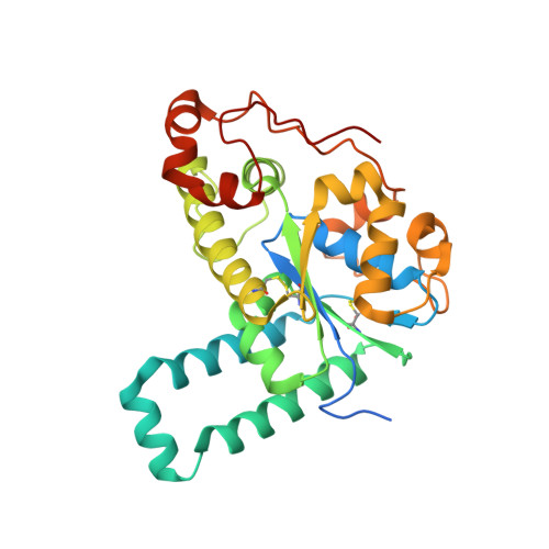
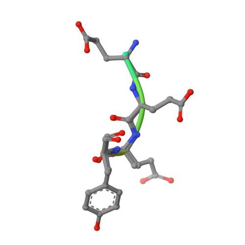
Crystal structure of human tyrosylprotein sulfotransferase-1 complexed with PAP and gastrin peptide


Experimental Data Snapshot

  • Method: X-RAY DIFFRACTION
  • Resolution: 2.31 Å
  • R-Value Free: 0.269 
  • R-Value Work: 0.241 
  • R-Value Observed: 0.242 

Starting Model: experimental
View more details

wwPDB Validation   3D Report Full Report


Ligand Structure Quality Assessment 


This is version 1.2 of the entry. See complete history


Literature

Structural basis for the broad substrate specificity of the human tyrosylprotein sulfotransferase-1.

Tanaka, S.Nishiyori, T.Kojo, H.Otsubo, R.Tsuruta, M.Kurogi, K.Liu, M.C.Suiko, M.Sakakibara, Y.Kakuta, Y.

(2017) Sci Rep 7: 8776-8776

  • DOI: https://doi.org/10.1038/s41598-017-07141-8
  • Primary Citation of Related Structures:  
    5WRI, 5WRJ

  • PubMed Abstract: 

    Tyrosylprotein sulfotransferases (TPSTs) are enzymes that catalyze post-translational tyrosine sulfation of proteins. In humans, there are only two TPST isoforms, designated TPST1 and TPST2. In a previous study, we reported the crystal structure of TPST2, which revealed the catalytic mechanism of the tyrosine sulfation reaction. However, detailed molecular mechanisms underlying how TPSTs catalyse a variety of substrate proteins with different efficiencies and how TPSTs catalyze the sulfation of multiple tyrosine residues in a substrate protein remain unresolved. Here, we report two crystal structures of the human TPST1 complexed with two substrate peptides that are catalysed by human TPST1 with significantly different efficiencies. The distinct binding modes found in the two complexes provide insight into the sulfation mechanism for these substrates. The present study provides valuable information describing the molecular mechanism of post-translational protein modifications catalysed by TPSTs.


  • Organizational Affiliation

    Department of Bioscience and Biotechnology, Faculty of Agriculture, Kyushu University, Hakozaki 6-10-1, Fukuoka, 812-8581, Japan.


Macromolecules
Find similar proteins by:  (by identity cutoff)  |  3D Structure
Entity ID: 1
MoleculeChains Sequence LengthOrganismDetailsImage
Protein-tyrosine sulfotransferase 1
A, B, C, D
303Homo sapiensMutation(s): 0 
Gene Names: TPST1
EC: 2.8.2.20
UniProt & NIH Common Fund Data Resources
Find proteins for O60507 (Homo sapiens)
Explore O60507 
Go to UniProtKB:  O60507
PHAROS:  O60507
GTEx:  ENSG00000169902 
Entity Groups  
Sequence Clusters30% Identity50% Identity70% Identity90% Identity95% Identity100% Identity
UniProt GroupO60507
Sequence Annotations
Expand
  • Reference Sequence

Find similar proteins by:  Sequence   |   3D Structure  

Entity ID: 2
MoleculeChains Sequence LengthOrganismDetailsImage
gastrin peptideE [auth F],
F [auth H],
G [auth J],
H [auth L]
12synthetic constructMutation(s): 0 
UniProt & NIH Common Fund Data Resources
Find proteins for P01350 (Homo sapiens)
Explore P01350 
Go to UniProtKB:  P01350
PHAROS:  P01350
GTEx:  ENSG00000184502 
Entity Groups  
Sequence Clusters30% Identity50% Identity70% Identity90% Identity95% Identity100% Identity
UniProt GroupP01350
Sequence Annotations
Expand
  • Reference Sequence
Experimental Data & Validation

Experimental Data

  • Method: X-RAY DIFFRACTION
  • Resolution: 2.31 Å
  • R-Value Free: 0.269 
  • R-Value Work: 0.241 
  • R-Value Observed: 0.242 
  • Space Group: P 1
Unit Cell:
Length ( Å )Angle ( ˚ )
a = 51.668α = 90.6
b = 93.263β = 93.39
c = 93.012γ = 103.39
Software Package:
Software NamePurpose
REFMACrefinement
HKL-2000data reduction
HKL-2000data scaling
MOLREPphasing

Structure Validation

View Full Validation Report


Ligand Structure Quality Assessment 


Entry History 

Deposition Data

Revision History  (Full details and data files)

  • Version 1.0: 2017-09-06
    Type: Initial release
  • Version 1.1: 2021-09-15
    Changes: Database references
  • Version 1.2: 2023-11-08
    Changes: Data collection, Refinement description