5V64 | pdb_00005v64

Crystal structure of macrocycles containing Abeta 15-21 (QKLV(PHI)FA) and Abeta 30-36 (AII(SAR)L(ORN)V)


Experimental Data Snapshot

  • Method: X-RAY DIFFRACTION
  • Resolution: 2.02 Å
  • R-Value Free: 
    0.244 (Depositor), 0.248 (DCC) 
  • R-Value Work: 
    0.195 (Depositor), 0.206 (DCC) 
  • R-Value Observed: 
    0.200 (Depositor) 

wwPDB Validation 3D Report Full Report

Validation slider image for 5V64

This is version 1.4 of the entry. See complete history

Literature

X-ray Crystallographic Structure of a Compact Dodecamer from a Peptide Derived from A beta 16-36.

Salveson, P.J.Spencer, R.K.Kreutzer, A.G.Nowick, J.S.

(2017) Org Lett 19: 3462-3465

  • DOI: https://doi.org/10.1021/acs.orglett.7b01445
  • Primary Citation Related Structures: 
    5V63, 5V64, 5V65

  • PubMed Abstract: 

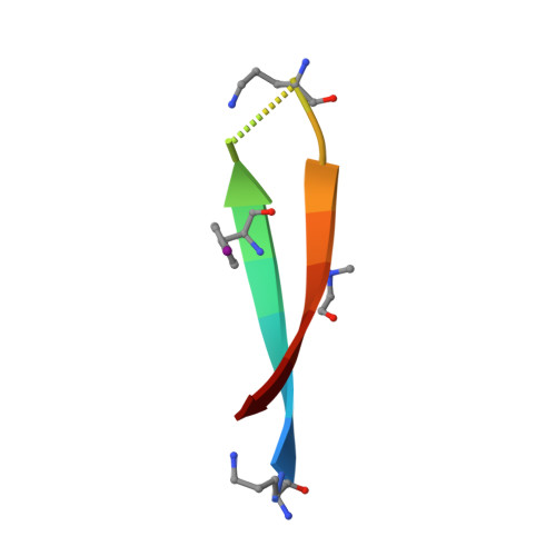
    The assembly of the β-amyloid peptide, Aβ, into soluble oligomers is associated with neurodegeneration in Alzheimer's disease. The Aβ oligomers are thought to be composed of β-hairpins. Here, the effect of shifting the residue pairing of the β-hairpins on the structures of the oligomers that form is explored through X-ray crystallography. Three residue pairings were investigated using constrained macrocyclic β-hairpins in which Aβ 30-36 is juxtaposed with Aβ 17-23 , Aβ 16-22 , and Aβ 15-21 . The Aβ 16-22 -Aβ 30-36 pairing forms a compact ball-shaped dodecamer composed of fused triangular trimers. This dodecamer may help explain the structures of the trimers and dodecamers formed by full-length Aβ.


  • Organizational Affiliation
    • Department of Chemistry, University of California, Irvine , Irvine, California 92697-2025, United States.

Macromolecule Content 

  • Total Structure Weight: 2.15 kDa 
  • Atom Count: 150 
  • Modeled Residue Count: 16 
  • Deposited Residue Count: 16 
  • Unique protein chains: 1

Macromolecules

Find similar proteins by:  Sequence   |   3D Structure  
Entity ID: 1
MoleculeChains  Sequence LengthOrganismDetailsImage
ORN-GLN-LYS-LEU-VAL-PHI-PHE-ALA-ORN-ALA-ILE-ILE-SAR-LEU-MET-VAL16synthetic constructMutation(s): 0 

Small Molecules

Modified Residues  3 Unique
IDChains TypeFormula2D DiagramParent
ORN
Query on ORN
A
L-PEPTIDE LINKINGC5 H12 N2 O2ALA
PHI
Query on PHI
A
L-PEPTIDE LINKINGC9 H10 I N O2PHE
SAR
Query on SAR
A
PEPTIDE LINKINGC3 H7 N O2GLY

Experimental Data & Validation

Experimental Data

  • Method: X-RAY DIFFRACTION
  • Resolution: 2.02 Å
  • R-Value Free:  0.244 (Depositor), 0.248 (DCC) 
  • R-Value Work:  0.195 (Depositor), 0.206 (DCC) 
  • R-Value Observed: 0.200 (Depositor) 
Space Group: P 21 21 2
Unit Cell:
Length ( Å )Angle ( ˚ )
a = 20.346α = 90
b = 22.029β = 90
c = 31.598γ = 90
Software Package:
Software NamePurpose
PHENIXrefinement
XDSdata reduction
Aimlessdata scaling
PHASERphasing

Structure Validation

View Full Validation Report


Entry History 

& Funding Information

Deposition Data


Funding OrganizationLocationGrant Number
National Institutes of Health/National Institute of General Medical Sciences (NIH/NIGMS)United StatesGM097562

Revision History  (Full details and data files)

  • Version 1.0: 2017-06-28
    Type: Initial release
  • Version 1.1: 2017-07-05
    Changes: Database references
  • Version 1.2: 2017-07-26
    Changes: Advisory, Author supporting evidence, Database references
  • Version 1.3: 2020-01-01
    Changes: Author supporting evidence
  • Version 1.4: 2025-04-02
    Changes: Data collection, Database references, Derived calculations, Structure summary