5V2I

Crystal structure of a mutant glycosylasparaginase (G172D) that causes the genetic disease Aspartylglucosaminuria


Experimental Data Snapshot

  • Method: X-RAY DIFFRACTION
  • Resolution: 1.83 Å
  • R-Value Free: 0.197 
  • R-Value Work: 0.156 
  • R-Value Observed: 0.158 

wwPDB Validation   3D Report Full Report


This is version 1.4 of the entry. See complete history


Literature

Crystal structure of a mutant glycosylasparaginase shedding light on aspartylglycosaminuria-causing mechanism as well as on hydrolysis of non-chitobiose substrate.

Pande, S.Lakshminarasimhan, D.Guo, H.C.

(2017) Mol Genet Metab 121: 150-156

  • DOI: https://doi.org/10.1016/j.ymgme.2017.04.008
  • Primary Citation of Related Structures:  
    5V2I

  • PubMed Abstract: 

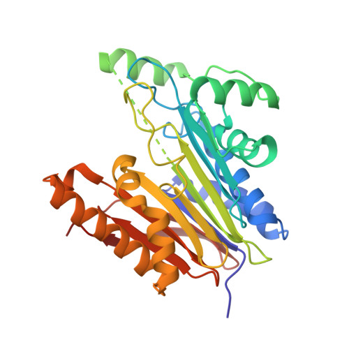
    Glycosylasparaginase (GA) is an amidase that cleaves Asn-linked glycoproteins in lysosomes. Deficiency of this enzyme causes accumulation of glycoasparagines in lysosomes of cells, resulting in a genetic condition called aspartylglycosaminuria (AGU). To better understand the mechanism of a disease-causing mutation with a single residue change from a glycine to an aspartic acid, we generated a model mutant enzyme at the corresponding position (named G172D mutant). Here we report a 1.8Å resolution crystal structure of mature G172D mutant and analyzed the reason behind its low hydrolase activity. Comparison of mature G172D and wildtype GA models reveals that the presence of Asp 172 near the catalytic site affects substrate catabolism in mature G172D, making it less efficient in substrate processing. Also recent studies suggest that GA is capable of processing substrates that lack a chitobiose (Glycan, N-acetylchiobios, NAcGlc) moiety, by its exo-hydrolase activity. The mechanism for this type of catalysis is not yet clear. l-Aspartic acid β-hydroxamate (β-AHA) is a non-chitobiose substrate that is known to interact with GA. To study the underlying mechanism of non-chitobiose substrate processing, we built a GA-β-AHA complex structure by comparing to a previously published G172D mutant precursor in complex with a β-AHA molecule. A hydrolysis mechanism of β-AHA by GA is proposed based on this complex model.


  • Organizational Affiliation

    Department of Biological Sciences, University of Massachusetts Lowell, 1 University Avenue, Lowell, MA 01854, USA.


Macromolecules
Find similar proteins by:  (by identity cutoff)  |  3D Structure
Entity ID: 1
MoleculeChains Sequence LengthOrganismDetailsImage
Glycosylasparaginase
A, B
295Elizabethkingia meningosepticaMutation(s): 1 
Gene Names: BES09_04975
UniProt
Find proteins for Q47898 (Elizabethkingia miricola)
Explore Q47898 
Go to UniProtKB:  Q47898
Entity Groups  
Sequence Clusters30% Identity50% Identity70% Identity90% Identity95% Identity100% Identity
UniProt GroupQ47898
Sequence Annotations
Expand
  • Reference Sequence
Experimental Data & Validation

Experimental Data

  • Method: X-RAY DIFFRACTION
  • Resolution: 1.83 Å
  • R-Value Free: 0.197 
  • R-Value Work: 0.156 
  • R-Value Observed: 0.158 
  • Space Group: P 1
Unit Cell:
Length ( Å )Angle ( ˚ )
a = 46.04α = 81.51
b = 52.77β = 90.15
c = 61.89γ = 105.93
Software Package:
Software NamePurpose
REFMACrefinement
iMOSFLMdata reduction
Aimlessdata scaling
MOLREPphasing

Structure Validation

View Full Validation Report


Entry History & Funding Information

Deposition Data


Funding OrganizationLocationGrant Number
National Institutes of Health/National Institute of Diabetes and Digestive and Kidney Disease (NIH/NIDDK)United StatesDK075294

Revision History  (Full details and data files)

  • Version 1.0: 2017-05-17
    Type: Initial release
  • Version 1.1: 2017-06-28
    Changes: Database references
  • Version 1.2: 2017-09-20
    Changes: Author supporting evidence, Refinement description
  • Version 1.3: 2019-12-25
    Changes: Author supporting evidence
  • Version 1.4: 2023-10-04
    Changes: Advisory, Data collection, Database references, Refinement description