5UJ7

Structure of the active form of human Origin Recognition Complex ATPase motor module, complex subunitS 1, 4, 5


Experimental Data Snapshot

  • Method: X-RAY DIFFRACTION
  • Resolution: 3.39 Å
  • R-Value Free: 0.281 
  • R-Value Work: 0.242 
  • R-Value Observed: 0.245 

wwPDB Validation   3D Report Full Report


This is version 1.3 of the entry. See complete history


Literature

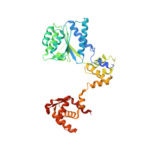
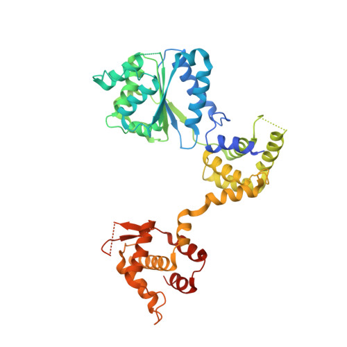
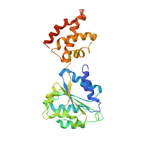
Structure of the active form of human Origin Recognition Complex and its ATPase motor module.

Tocilj, A.On, K.F.Yuan, Z.Sun, J.Elkayam, E.Li, H.Stillman, B.Joshua-Tor, L.

(2017) Elife 6

  • DOI: https://doi.org/10.7554/eLife.20818
  • Primary Citation of Related Structures:  
    5UJ7, 5UJ8, 5UJM

  • PubMed Abstract: 

    Binding of the Origin Recognition Complex (ORC) to origins of replication marks the first step in the initiation of replication of the genome in all eukaryotic cells. Here, we report the structure of the active form of human ORC determined by X-ray crystallography and cryo-electron microscopy. The complex is composed of an ORC1/4/5 motor module lobe in an organization reminiscent of the DNA polymerase clamp loader complexes. A second lobe contains the ORC2/3 subunits. The complex is organized as a double-layered shallow corkscrew, with the AAA+ and AAA+-like domains forming one layer, and the winged-helix domains (WHDs) forming a top layer. CDC6 fits easily between ORC1 and ORC2, completing the ring and the DNA-binding channel, forming an additional ATP hydrolysis site. Analysis of the ATPase activity of the complex provides a basis for understanding ORC activity as well as molecular defects observed in Meier-Gorlin Syndrome mutations.


  • Organizational Affiliation

    W. M. Keck Structural Biology Laboratory, Cold Spring Harbor, New York, United States.


Macromolecules
Find similar proteins by:  (by identity cutoff)  |  3D Structure
Entity ID: 1
MoleculeChains Sequence LengthOrganismDetailsImage
Origin recognition complex subunit 1A,
D [auth B]
391Homo sapiensMutation(s): 0 
Gene Names: ORC1ORC1LPARC1
UniProt & NIH Common Fund Data Resources
Find proteins for Q13415 (Homo sapiens)
Explore Q13415 
Go to UniProtKB:  Q13415
PHAROS:  Q13415
GTEx:  ENSG00000085840 
Entity Groups  
Sequence Clusters30% Identity50% Identity70% Identity90% Identity95% Identity100% Identity
UniProt GroupQ13415
Sequence Annotations
Expand
  • Reference Sequence
Find similar proteins by:  (by identity cutoff)  |  3D Structure
Entity ID: 2
MoleculeChains Sequence LengthOrganismDetailsImage
Origin recognition complex subunit 4B [auth C],
E [auth D]
436Homo sapiensMutation(s): 0 
Gene Names: ORC4ORC4L
UniProt & NIH Common Fund Data Resources
Find proteins for O43929 (Homo sapiens)
Explore O43929 
Go to UniProtKB:  O43929
PHAROS:  O43929
GTEx:  ENSG00000115947 
Entity Groups  
Sequence Clusters30% Identity50% Identity70% Identity90% Identity95% Identity100% Identity
UniProt GroupO43929
Sequence Annotations
Expand
  • Reference Sequence
Find similar proteins by:  (by identity cutoff)  |  3D Structure
Entity ID: 3
MoleculeChains Sequence LengthOrganismDetailsImage
Origin recognition complex subunit 5C [auth E],
F
284Homo sapiensMutation(s): 0 
Gene Names: ORC5ORC5L
UniProt & NIH Common Fund Data Resources
Find proteins for O43913 (Homo sapiens)
Explore O43913 
Go to UniProtKB:  O43913
PHAROS:  O43913
GTEx:  ENSG00000164815 
Entity Groups  
Sequence Clusters30% Identity50% Identity70% Identity90% Identity95% Identity100% Identity
UniProt GroupO43913
Sequence Annotations
Expand
  • Reference Sequence
Small Molecules
Ligands 3 Unique
IDChains Name / Formula / InChI Key2D Diagram3D Interactions
ATP
Query on ATP

Download Ideal Coordinates CCD File 
G [auth A]
I [auth C]
K [auth E]
M [auth B]
O [auth D]
G [auth A],
I [auth C],
K [auth E],
M [auth B],
O [auth D],
Q [auth F]
ADENOSINE-5'-TRIPHOSPHATE
C10 H16 N5 O13 P3
ZKHQWZAMYRWXGA-KQYNXXCUSA-N
K
Query on K

Download Ideal Coordinates CCD File 
L [auth E],
R [auth F]
POTASSIUM ION
K
NPYPAHLBTDXSSS-UHFFFAOYSA-N
MG
Query on MG

Download Ideal Coordinates CCD File 
H [auth A],
J [auth C],
N [auth B],
P [auth D]
MAGNESIUM ION
Mg
JLVVSXFLKOJNIY-UHFFFAOYSA-N
Experimental Data & Validation

Experimental Data

  • Method: X-RAY DIFFRACTION
  • Resolution: 3.39 Å
  • R-Value Free: 0.281 
  • R-Value Work: 0.242 
  • R-Value Observed: 0.245 
  • Space Group: P 1 21 1
Unit Cell:
Length ( Å )Angle ( ˚ )
a = 120.887α = 90
b = 81.141β = 97.25
c = 151.947γ = 90
Software Package:
Software NamePurpose
PHENIXrefinement
XDSdata reduction
Aimlessdata scaling
PHASERphasing

Structure Validation

View Full Validation Report


Entry History & Funding Information

Deposition Data


Funding OrganizationLocationGrant Number
Howard Hughes Medical Institute (HHMI)United States--

Revision History  (Full details and data files)

  • Version 1.0: 2017-02-08
    Type: Initial release
  • Version 1.1: 2017-09-20
    Changes: Author supporting evidence, Derived calculations
  • Version 1.2: 2019-11-20
    Changes: Author supporting evidence
  • Version 1.3: 2023-10-04
    Changes: Data collection, Database references, Derived calculations, Refinement description