5OIU

Crystal structure of PilF type IV pilus assembly ATPase from Thermus thermophilus


Experimental Data Snapshot

  • Method: X-RAY DIFFRACTION
  • Resolution: 2.44 Å
  • R-Value Free: 0.246 
  • R-Value Work: 0.190 
  • R-Value Observed: 0.193 

wwPDB Validation   3D Report Full Report


Ligand Structure Quality Assessment 


This is version 1.2 of the entry. See complete history


Literature

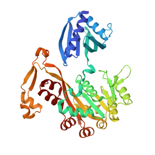
Structural cycle of the Thermus thermophilus PilF ATPase: the powering of type IVa pilus assembly.

Collins, R.Karuppiah, V.Siebert, C.A.Dajani, R.Thistlethwaite, A.Derrick, J.P.

(2018) Sci Rep 8: 14022-14022

  • DOI: https://doi.org/10.1038/s41598-018-32218-3
  • Primary Citation of Related Structures:  
    5OIU, 6EJF, 6F8L

  • PubMed Abstract: 

    Type IV pili are responsible for a diverse range of functions, including twitching motility and cell adhesion. Assembly of the pilus fiber is driven by a cytoplasmic ATPase: it interacts with an inner membrane complex of biogenesis proteins which, in turn, bind to nascent pilin subunits and mediate fiber assembly. Here we report the structural characterization of the PilF TFP assembly ATPase from Thermus thermophilus. The crystal structure of a recombinant C-terminal fragment of PilF revealed bound, unhydrolysed ATP, although the full length complex was enzymatically active. 3D reconstructions were carried out by single particle cryoelectron microscopy for full length apoprotein PilF and in complex with AMPPNP. The structure forms an hourglass-like shape, with the ATPase domains in one half and the N1 domains in the second half which, we propose, interact with the other pilus biogenesis components. Molecular models for both forms were generated: binding of AMPPNP causes an upward shift of the N1 domains towards the ATPase domains of ~8 Å. We advocate a model in which ATP hydrolysis is linked to displacement of the N1 domains which is associated with lifting pilin subunits out of the inner membrane, and provide the activation energy needed to form the pilus fiber.


  • Organizational Affiliation

    School of Biological Sciences, Faculty of Biology, Medicine and Health, Manchester Academic Health Science Centre, The University of Manchester, Oxford Road, Manchester, UK.


Macromolecules
Find similar proteins by:  (by identity cutoff)  |  3D Structure
Entity ID: 1
MoleculeChains Sequence LengthOrganismDetailsImage
Type IV pilus assembly protein PilF
A, B, C, D, E
A, B, C, D, E, F
390Thermus thermophilus HB8Mutation(s): 0 
Gene Names: TTHA0364
UniProt
Find proteins for Q5SLC9 (Thermus thermophilus (strain ATCC 27634 / DSM 579 / HB8))
Explore Q5SLC9 
Go to UniProtKB:  Q5SLC9
Entity Groups  
Sequence Clusters30% Identity50% Identity70% Identity90% Identity95% Identity100% Identity
UniProt GroupQ5SLC9
Sequence Annotations
Expand
  • Reference Sequence
Small Molecules
Ligands 4 Unique
IDChains Name / Formula / InChI Key2D Diagram3D Interactions
ATP
Query on ATP

Download Ideal Coordinates CCD File 
CA [auth E]
G [auth A]
GA [auth F]
L [auth B]
Q [auth C]
CA [auth E],
G [auth A],
GA [auth F],
L [auth B],
Q [auth C],
W [auth D]
ADENOSINE-5'-TRIPHOSPHATE
C10 H16 N5 O13 P3
ZKHQWZAMYRWXGA-KQYNXXCUSA-N
ZN
Query on ZN

Download Ideal Coordinates CCD File 
EA [auth E]
I [auth A]
IA [auth F]
N [auth B]
S [auth C]
EA [auth E],
I [auth A],
IA [auth F],
N [auth B],
S [auth C],
Y [auth D]
ZINC ION
Zn
PTFCDOFLOPIGGS-UHFFFAOYSA-N
CL
Query on CL

Download Ideal Coordinates CCD File 
AA [auth D]
BA [auth D]
FA [auth E]
J [auth A]
JA [auth F]
AA [auth D],
BA [auth D],
FA [auth E],
J [auth A],
JA [auth F],
K [auth A],
O [auth B],
P [auth B],
T [auth C],
U [auth C],
V [auth C],
Z [auth D]
CHLORIDE ION
Cl
VEXZGXHMUGYJMC-UHFFFAOYSA-M
MG
Query on MG

Download Ideal Coordinates CCD File 
DA [auth E]
H [auth A]
HA [auth F]
M [auth B]
R [auth C]
DA [auth E],
H [auth A],
HA [auth F],
M [auth B],
R [auth C],
X [auth D]
MAGNESIUM ION
Mg
JLVVSXFLKOJNIY-UHFFFAOYSA-N
Experimental Data & Validation

Experimental Data

  • Method: X-RAY DIFFRACTION
  • Resolution: 2.44 Å
  • R-Value Free: 0.246 
  • R-Value Work: 0.190 
  • R-Value Observed: 0.193 
  • Space Group: C 1 2 1
Unit Cell:
Length ( Å )Angle ( ˚ )
a = 220.39α = 90
b = 107.66β = 112.95
c = 137.83γ = 90
Software Package:
Software NamePurpose
PHENIXrefinement
XDSdata reduction
XSCALEdata scaling
PHASERphasing
PHENIXmodel building

Structure Validation

View Full Validation Report


Ligand Structure Quality Assessment 


Entry History & Funding Information

Deposition Data


Funding OrganizationLocationGrant Number
Wellcome TrustUnited Kingdom093388/Z/10/Z

Revision History  (Full details and data files)

  • Version 1.0: 2018-08-08
    Type: Initial release
  • Version 1.1: 2019-02-20
    Changes: Data collection, Database references
  • Version 1.2: 2024-01-17
    Changes: Data collection, Database references, Derived calculations, Refinement description