5N2V

Changes in conformational equilibria regulate the activity of the Dcp2 decapping enzyme


Experimental Data Snapshot

  • Method: X-RAY DIFFRACTION
  • Resolution: 3.10 Å
  • R-Value Free: 0.287 
  • R-Value Work: 0.239 
  • R-Value Observed: 0.241 

wwPDB Validation   3D Report Full Report


Ligand Structure Quality Assessment 


This is version 2.0 of the entry. See complete history


Literature

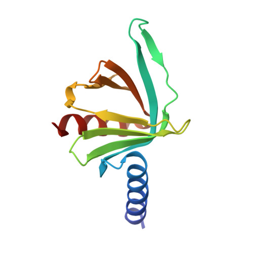
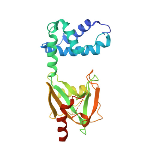
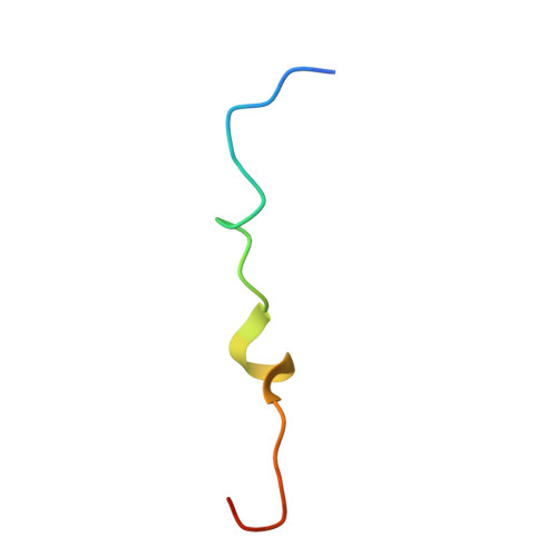
Changes in conformational equilibria regulate the activity of the Dcp2 decapping enzyme.

Wurm, J.P.Holdermann, I.Overbeck, J.H.Mayer, P.H.O.Sprangers, R.

(2017) Proc Natl Acad Sci U S A 114: 6034-6039

  • DOI: https://doi.org/10.1073/pnas.1704496114
  • Primary Citation of Related Structures:  
    5N2V

  • PubMed Abstract: 

    Crystal structures of enzymes are indispensable to understanding their mechanisms on a molecular level. It, however, remains challenging to determine which structures are adopted in solution, especially for dynamic complexes. Here, we study the bilobed decapping enzyme Dcp2 that removes the 5' cap structure from eukaryotic mRNA and thereby efficiently terminates gene expression. The numerous Dcp2 structures can be grouped into six states where the domain orientation between the catalytic and regulatory domains significantly differs. Despite this wealth of structural information it is not possible to correlate these states with the catalytic cycle or the activity of the enzyme. Using methyl transverse relaxation-optimized NMR spectroscopy, we demonstrate that only three of the six domain orientations are present in solution, where Dcp2 adopts an open, a closed, or a catalytically active state. We show how mRNA substrate and the activator proteins Dcp1 and Edc1 influence the dynamic equilibria between these states and how this modulates catalytic activity. Importantly, the active state of the complex is only stably formed in the presence of both activators and the mRNA substrate or the m7GDP decapping product, which we rationalize based on a crystal structure of the Dcp1:Dcp2:Edc1:m7GDP complex. Interestingly, we find that the activating mechanisms in Dcp2 also result in a shift of the substrate specificity from bacterial to eukaryotic mRNA.


  • Organizational Affiliation

    Max Planck Institute for Developmental Biology, 72076 Tuebingen, Germany.


Macromolecules
Find similar proteins by:  (by identity cutoff)  |  3D Structure
Entity ID: 1
MoleculeChains Sequence LengthOrganismDetailsImage
mRNA-decapping enzyme subunit 1
A, D
129Schizosaccharomyces pombeMutation(s): 0 
Gene Names: dcp1SPBC3B9.21
UniProt
Find proteins for Q9P805 (Schizosaccharomyces pombe (strain 972 / ATCC 24843))
Explore Q9P805 
Go to UniProtKB:  Q9P805
Entity Groups  
Sequence Clusters30% Identity50% Identity70% Identity90% Identity95% Identity100% Identity
UniProt GroupQ9P805
Sequence Annotations
Expand
  • Reference Sequence
Find similar proteins by:  (by identity cutoff)  |  3D Structure
Entity ID: 2
MoleculeChains Sequence LengthOrganismDetailsImage
mRNA decapping complex subunit 2
B, E
245Schizosaccharomyces pombeMutation(s): 0 
Gene Names: dcp2SPAC19A8.12
EC: 3
UniProt
Find proteins for O13828 (Schizosaccharomyces pombe (strain 972 / ATCC 24843))
Explore O13828 
Go to UniProtKB:  O13828
Entity Groups  
Sequence Clusters30% Identity50% Identity70% Identity90% Identity95% Identity100% Identity
UniProt GroupO13828
Sequence Annotations
Expand
  • Reference Sequence
Find similar proteins by:  (by identity cutoff)  |  3D Structure
Entity ID: 3
MoleculeChains Sequence LengthOrganismDetailsImage
Edc1
C, F
26Schizosaccharomyces pombeMutation(s): 0 
UniProt
Find proteins for Q10108 (Schizosaccharomyces pombe (strain 972 / ATCC 24843))
Explore Q10108 
Go to UniProtKB:  Q10108
Entity Groups  
Sequence Clusters30% Identity50% Identity70% Identity90% Identity95% Identity100% Identity
UniProt GroupQ10108
Sequence Annotations
Expand
  • Reference Sequence
Experimental Data & Validation

Experimental Data

  • Method: X-RAY DIFFRACTION
  • Resolution: 3.10 Å
  • R-Value Free: 0.287 
  • R-Value Work: 0.239 
  • R-Value Observed: 0.241 
  • Space Group: P 1 21 1
Unit Cell:
Length ( Å )Angle ( ˚ )
a = 63.841α = 90
b = 76.522β = 102.83
c = 89.526γ = 90
Software Package:
Software NamePurpose
PHENIXrefinement
XDSdata reduction
SCALAdata scaling
PHASERphasing

Structure Validation

View Full Validation Report


Ligand Structure Quality Assessment 


Entry History & Funding Information

Deposition Data


Funding OrganizationLocationGrant Number
EUERC-616052

Revision History  (Full details and data files)

  • Version 1.0: 2017-05-24
    Type: Initial release
  • Version 1.1: 2017-06-07
    Changes: Database references
  • Version 1.2: 2017-06-14
    Changes: Database references
  • Version 1.3: 2024-01-17
    Changes: Data collection, Database references, Derived calculations, Refinement description
  • Version 2.0: 2024-02-28
    Changes: Atomic model, Data collection, Derived calculations, Non-polymer description, Structure summary