5MSE | pdb_00005mse

GFP nuclear transport receptor mimic 3B8


Experimental Data Snapshot

  • Method: X-RAY DIFFRACTION
  • Resolution: 1.66 Å
  • R-Value Free: 
    0.206 (Depositor), 0.216 (DCC) 
  • R-Value Work: 
    0.174 (Depositor), 0.186 (DCC) 
  • R-Value Observed: 
    0.175 (Depositor) 

Starting Model: experimental
View more details

wwPDB Validation 3D Report Full Report

Validation slider image for 5MSE

This is version 1.4 of the entry. See complete history

Literature

Surface Properties Determining Passage Rates of Proteins through Nuclear Pores.

Frey, S.Rees, R.Schunemann, J.Ng, S.C.Funfgeld, K.Huyton, T.Gorlich, D.

(2018) Cell 174: 202-217.e9

  • DOI: https://doi.org/10.1016/j.cell.2018.05.045
  • Primary Citation Related Structures: 
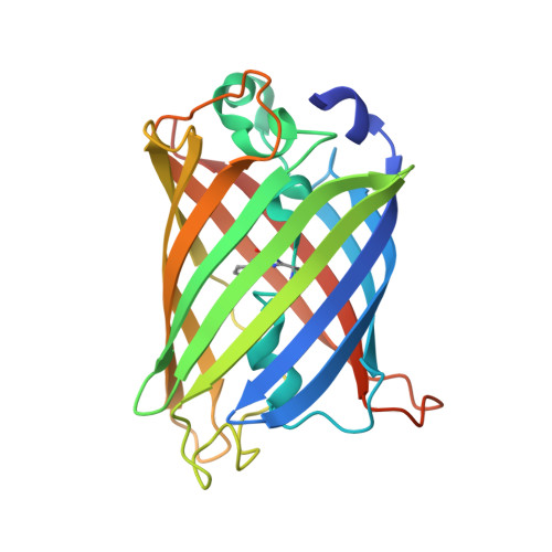
    5MSE

  • PubMed Abstract: 

    Nuclear pore complexes (NPCs) conduct nucleocytoplasmic transport through an FG domain-controlled barrier. We now explore how surface-features of a mobile species determine its NPC passage rate. Negative charges and lysines impede passage. Hydrophobic residues, certain polar residues (Cys, His), and, surprisingly, charged arginines have striking translocation-promoting effects. Favorable cation-π interactions between arginines and FG-phenylalanines may explain this apparent paradox. Application of these principles to redesign the surface of GFP resulted in variants that show a wide span of transit rates, ranging from 35-fold slower than wild-type to ∼500 times faster, with the latter outpacing even naturally occurring nuclear transport receptors (NTRs). The structure of a fast and particularly FG-specific GFP NTR variant illustrates how NTRs can expose multiple regions for binding hydrophobic FG motifs while evading non-specific aggregation. Finally, we document that even for NTR-mediated transport, the surface-properties of the "passively carried" cargo can strikingly affect the translocation rate.


  • Organizational Affiliation
    • Department of Cellular Logistics, Max Planck Institute for Biophysical Chemistry, Göttingen, Germany.

Macromolecule Content 

  • Total Structure Weight: 110.86 kDa 
  • Atom Count: 8,179 
  • Modeled Residue Count: 899 
  • Deposited Residue Count: 952 
  • Unique protein chains: 1

Macromolecules

Find similar proteins by:|  3D Structure
Entity ID: 1
MoleculeChains  Sequence LengthOrganismDetailsImage
Green fluorescent proteinA,
B [auth C],
C [auth D],
D [auth B]
238Aequorea victoriaMutation(s): 46 
Gene Names: GFP
UniProt
Find proteins for P42212 (Aequorea victoria)
Explore P42212 
Go to UniProtKB:  P42212
Entity Groups
Sequence Clusters30% Identity50% Identity70% Identity90% Identity95% Identity100% Identity
UniProt GroupP42212
Sequence Annotations
Expand
Reference Sequence

Small Molecules

Ligands 2 Unique
IDChains Name / Formula / InChI Key2D Diagram3D Interactions
IMD

Query on IMD


Download:Ideal Coordinates CCD File
G [auth A],
H [auth A],
K [auth C],
P [auth B]
IMIDAZOLE
C3 H5 N2
RAXXELZNTBOGNW-UHFFFAOYSA-O
NA

Query on NA


Download:Ideal Coordinates CCD File
E [auth A]
F [auth A]
I [auth C]
J [auth C]
L [auth D]
E [auth A],
F [auth A],
I [auth C],
J [auth C],
L [auth D],
M [auth D],
N [auth B],
O [auth B]
SODIUM ION
Na
FKNQFGJONOIPTF-UHFFFAOYSA-N
Modified Residues  1 Unique
IDChains TypeFormula2D DiagramParent
CRO
Query on CRO
A,
B [auth C],
C [auth D],
D [auth B]
L-PEPTIDE LINKINGC15 H17 N3 O5THR, TYR, GLY

Experimental Data & Validation

Experimental Data

  • Method: X-RAY DIFFRACTION
  • Resolution: 1.66 Å
  • R-Value Free:  0.206 (Depositor), 0.216 (DCC) 
  • R-Value Work:  0.174 (Depositor), 0.186 (DCC) 
  • R-Value Observed: 0.175 (Depositor) 
Space Group: P 1 21 1
Unit Cell:
Length ( Å )Angle ( ˚ )
a = 73.401α = 90
b = 73.398β = 93.73
c = 93.927γ = 90
Software Package:
Software NamePurpose
REFMACrefinement
XDSdata reduction
Aimlessdata scaling
PHASERphasing

Structure Validation

View Full Validation Report


Entry History 

Deposition Data

Revision History  (Full details and data files)

  • Version 1.0: 2018-05-02
    Type: Initial release
  • Version 1.1: 2018-06-13
    Changes: Data collection, Database references, Structure summary
  • Version 1.2: 2019-05-15
    Changes: Data collection, Database references
  • Version 1.3: 2024-01-17
    Changes: Data collection, Database references, Derived calculations, Refinement description
  • Version 1.4: 2024-11-20
    Changes: Structure summary