


Crystal structure of SmAP (LSm) protein from Methanococcus vannielii in complex with URIDINE-5'-MONOPHOSPHATE
Nikulin, A.D., Lekontseva, N.V., Tishchenko, S.V., Kravchenko, O.V.To be published.
Experimental Data Snapshot
Starting Model: experimental
View more details
Entity ID: 1 | |||||
|---|---|---|---|---|---|
| Molecule | Chains | Sequence Length | Organism | Details | Image |
| Like-Sm ribonucleoprotein core | 72 | Methanococcus vannielii | Mutation(s): 0 Gene Names: Mevan_0470 | 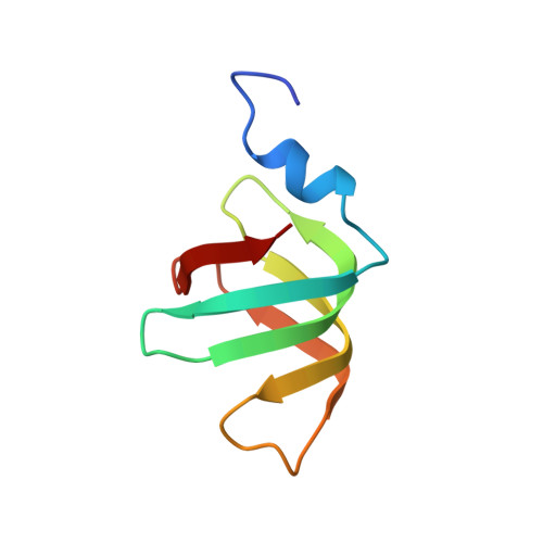 | |
UniProt | |||||
Entity Groups | |||||
| Sequence Clusters | 30% Identity50% Identity70% Identity90% Identity95% Identity100% Identity | ||||
| UniProt Group | A6UPF5 | ||||
Sequence AnnotationsExpand | |||||
Reference Sequence | |||||
| Ligands 2 Unique | |||||
|---|---|---|---|---|---|
| ID | Chains | Name / Formula / InChI Key | 2D Diagram | 3D Interactions | |
| U5P Download:Ideal Coordinates CCD File | AB [auth R] BB [auth T] CA [auth A] CB [auth U] DA [auth B] | URIDINE-5'-MONOPHOSPHATE C9 H13 N2 O9 P DJJCXFVJDGTHFX-XVFCMESISA-N |  | ||
| PG4 Download:Ideal Coordinates CCD File | EA [auth B] GB [auth W] HA [auth D] JA [auth E] KB [auth Z] | TETRAETHYLENE GLYCOL C8 H18 O5 UWHCKJMYHZGTIT-UHFFFAOYSA-N |  | ||
| Length ( Å ) | Angle ( ˚ ) |
|---|---|
| a = 91.015 | α = 90 |
| b = 97.869 | β = 103.88 |
| c = 123.448 | γ = 90 |
| Software Name | Purpose |
|---|---|
| PHENIX | refinement |
| XDS | data reduction |
| XSCALE | data scaling |
| PHASER | phasing |
| Funding Organization | Location | Grant Number |
|---|---|---|
| Russian Scientific Foundation | Russian Federation | 14-14-00496 |


