5MGA | pdb_00005mga

Structure of the Cpf1 endonuclease R-loop complex after DNA cleavage


Experimental Data Snapshot

  • Method: X-RAY DIFFRACTION
  • Resolution: 3.00 Å
  • R-Value Free: 
    0.265 (Depositor), 0.260 (DCC) 
  • R-Value Work: 
    0.242 (Depositor), 0.241 (DCC) 
  • R-Value Observed: 
    0.243 (Depositor) 

wwPDB Validation 3D Report Full Report

Validation slider image for 5MGA

This is version 1.3 of the entry. See complete history

Literature

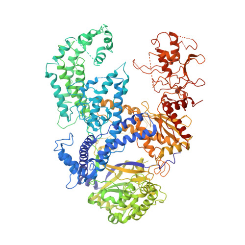
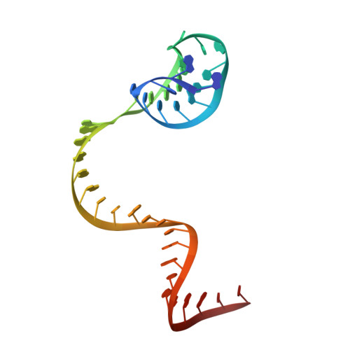
Structure of the Cpf1 endonuclease R-loop complex after target DNA cleavage.

Stella, S.Alcon, P.Montoya, G.

(2017) Nature 546: 559-563

  • DOI: https://doi.org/10.1038/nature22398
  • Primary Citation Related Structures: 
    5MGA

  • PubMed Abstract: 

    Cpf1 is an RNA-guided endonuclease that is emerging as a powerful genome-editing tool. Here we provide insight into its DNA-targeting mechanism by determining the structure of Francisella novicida Cpf1 with the triple-stranded R-loop generated after DNA cleavage. The structure reveals the machinery involved in DNA unwinding to form a CRISPR RNA (crRNA)-DNA hybrid and a displaced DNA strand. The protospacer adjacent motif (PAM) is recognized by the PAM-interacting domain. The loop-lysine helix-loop motif in this domain contains three conserved lysine residues that are inserted in a dentate manner into the double-stranded DNA. Unzipping of the double-stranded DNA occurs in a cleft arranged by acidic and hydrophobic residues facilitating the crRNA-DNA hybrid formation. The PAM single-stranded DNA is funnelled towards the nuclease site through a mixed hydrophobic and basic cavity. In this catalytic conformation, the PAM-interacting domain and the helix-loop-helix motif in the REC1 domain adopt a 'rail' shape and 'flap-on' conformations, respectively, channelling the PAM strand into the cavity. A steric barrier between the RuvC-II and REC1 domains forms the 'septum', separating the displaced PAM strand and the crRNA-DNA hybrid, avoiding DNA re-annealing. Mutations in key residues reveal a mechanism linking the PAM and DNA nuclease sites. Analysis of the Cpf1 structures proposes a singular working model of RNA-guided DNA cleavage, suggesting new avenues for redesign of Cpf1.


  • Organizational Affiliation
    • Protein Structure &Function Programme, Macromolecular Crystallography Group, Novo Nordisk Foundation Center for Protein Research, Faculty of Health and Medical Sciences, University of Copenhagen, Blegdamsvej 3B, Copenhagen 2200, Denmark.

Macromolecule Content 

  • Total Structure Weight: 179.33 kDa 
  • Atom Count: 11,351 
  • Modeled Residue Count: 1,260 
  • Deposited Residue Count: 1,401 
  • Unique protein chains: 1
  • Unique nucleic acid chains: 3

Macromolecules


Find similar proteins by:|  3D Structure
Entity ID: 1
MoleculeChains  Sequence LengthOrganismDetailsImage
CRISPR-associated endonuclease Cpf11,323Francisella tularensis subsp. novicida U112Mutation(s): 0 
Gene Names: cpf1FTN_1397
EC: 3.1 (PDB Primary Data), 4.6.1.22 (UniProt), 3.1.21.1 (UniProt)
UniProt
Find proteins for A0Q7Q2 (Francisella tularensis subsp. novicida (strain ATCC 15482 / CCUG 33449 / U112))
Explore A0Q7Q2 
Go to UniProtKB:  A0Q7Q2
Entity Groups
Sequence Clusters30% Identity50% Identity70% Identity90% Identity95% Identity100% Identity
UniProt GroupA0Q7Q2
Sequence Annotations
Expand
Reference Sequence
Find similar nucleic acids by:  (by identity cutoff) 
Entity ID: 2
MoleculeChains LengthOrganismImage
RNA (40-MER)40Francisella tularensis subsp. novicida U112
Sequence Annotations
Expand
Reference Sequence
Find similar nucleic acids by:  (by identity cutoff) 
Entity ID: 3
MoleculeChains LengthOrganismImage
DNA (26-MER)26Francisella tularensis subsp. novicida U112
Sequence Annotations
Expand
Reference Sequence
Find similar nucleic acids by:  Sequence
Entity ID: 4
MoleculeChains LengthOrganismImage
DNA (5'-D(P*CP*GP*TP*TP*AP*GP*AP*GP*AP*AP*GP*T)-3')12Francisella tularensis subsp. novicida U112
Sequence Annotations
Expand
Reference Sequence

Experimental Data & Validation

Experimental Data

  • Method: X-RAY DIFFRACTION
  • Resolution: 3.00 Å
  • R-Value Free:  0.265 (Depositor), 0.260 (DCC) 
  • R-Value Work:  0.242 (Depositor), 0.241 (DCC) 
  • R-Value Observed: 0.243 (Depositor) 
Space Group: C 2 2 21
Unit Cell:
Length ( Å )Angle ( ˚ )
a = 85.223α = 90
b = 137.652β = 90
c = 320.513γ = 90
Software Package:
Software NamePurpose
REFMACrefinement
XDSdata reduction
Aimlessdata scaling
PHASERphasing

Structure Validation

View Full Validation Report


Entry History 

& Funding Information

Deposition Data


Funding OrganizationLocationGrant Number
Novo Nordisk FoundationDenmarkNNF14CC0001

Revision History  (Full details and data files)

  • Version 1.0: 2017-06-21
    Type: Initial release
  • Version 1.1: 2017-06-28
    Changes: Database references
  • Version 1.2: 2018-10-24
    Changes: Advisory, Data collection, Derived calculations
  • Version 1.3: 2024-05-08
    Changes: Data collection, Database references