Proteins evolve on the edge of supramolecular self-assembly.
Garcia-Seisdedos, H., Empereur-Mot, C., Elad, N., Levy, E.D.(2017) Nature 548: 244-247
- PubMed: 28783726 Search on PubMed
- DOI: https://doi.org/10.1038/nature23320
- Primary Citation Related Structures:
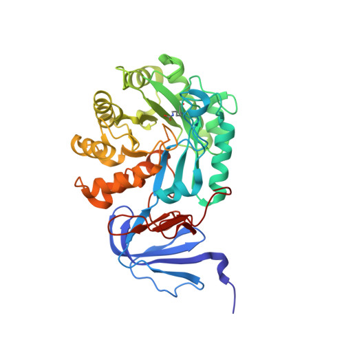
5LP3 - PubMed Abstract:
The self-association of proteins into symmetric complexes is ubiquitous in all kingdoms of life. Symmetric complexes possess unique geometric and functional properties, but their internal symmetry can pose a risk. In sickle-cell disease, the symmetry of haemoglobin exacerbates the effect of a mutation, triggering assembly into harmful fibrils. Here we examine the universality of this mechanism and its relation to protein structure geometry. We introduced point mutations solely designed to increase surface hydrophobicity among 12 distinct symmetric complexes from Escherichia coli. Notably, all responded by forming supramolecular assemblies in vitro, as well as in vivo upon heterologous expression in Saccharomyces cerevisiae. Remarkably, in four cases, micrometre-long fibrils formed in vivo in response to a single point mutation. Biophysical measurements and electron microscopy revealed that mutants self-assembled in their folded states and so were not amyloid-like. Structural examination of 73 mutants identified supramolecular assembly hot spots predictable by geometry. A subsequent structural analysis of 7,471 symmetric complexes showed that geometric hot spots were buffered chemically by hydrophilic residues, suggesting a mechanism preventing mis-assembly of these regions. Thus, point mutations can frequently trigger folded proteins to self-assemble into higher-order structures. This potential is counterbalanced by negative selection and can be exploited to design nanomaterials in living cells.
- Department of Structural Biology, Weizmann Institute of Science, Rehovot 7610001, Israel.
Organizational Affiliation: