5KMG

Near-atomic cryo-EM structure of PRC1 bound to the microtubule


Experimental Data Snapshot

  • Method: ELECTRON MICROSCOPY
  • Resolution: 3.50 Å
  • Aggregation State: HELICAL ARRAY 
  • Reconstruction Method: HELICAL 

wwPDB Validation   3D Report Full Report


This is version 1.4 of the entry. See complete history


Literature

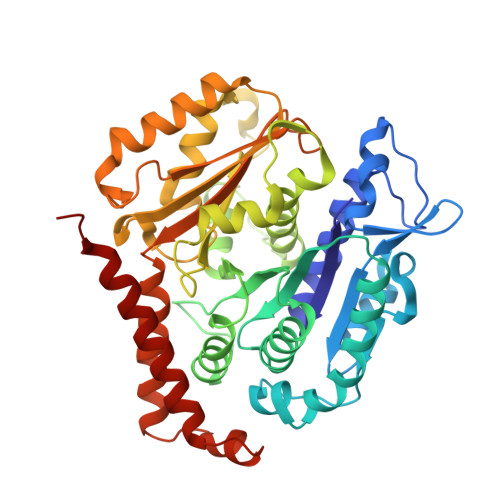
Near-atomic cryo-EM structure of PRC1 bound to the microtubule.

Kellogg, E.H.Howes, S.Ti, S.C.Ramirez-Aportela, E.Kapoor, T.M.Chacon, P.Nogales, E.

(2016) Proc Natl Acad Sci U S A 113: 9430-9439

  • DOI: 10.1073/pnas.1609903113
  • Primary Citation of Related Structures:  
    5KMG

  • PubMed Abstract: 
  • Proteins that associate with microtubules (MTs) are crucial to generate MT arrays and establish different cellular architectures. One example is PRC1 (protein regulator of cytokinesis 1), which cross-links antiparallel MTs and is essential for the completion of mitosis and cytokinesis ...

    Proteins that associate with microtubules (MTs) are crucial to generate MT arrays and establish different cellular architectures. One example is PRC1 (protein regulator of cytokinesis 1), which cross-links antiparallel MTs and is essential for the completion of mitosis and cytokinesis. Here we describe a 4-Å-resolution cryo-EM structure of monomeric PRC1 bound to MTs. Residues in the spectrin domain of PRC1 contacting the MT are highly conserved and interact with the same pocket recognized by kinesin. We additionally found that PRC1 promotes MT assembly even in the presence of the MT stabilizer taxol. Interestingly, the angle of the spectrin domain on the MT surface corresponds to the previously observed cross-bridge angle between MTs cross-linked by full-length, dimeric PRC1. This finding, together with molecular dynamic simulations describing the intrinsic flexibility of PRC1, suggests that the MT-spectrin domain interface determines the geometry of the MT arrays cross-linked by PRC1.


    Organizational Affiliation

    Molecular Biophysics and Integrative Bioimaging Division, Lawrence Berkeley National Laboratory, Berkeley, CA 94720; Howard Hughes Medical Institute, University of California, Berkeley, CA 94720; California Institute for Quantitative Biosciences, University of California, Berkeley, CA 94720; Department of Molecular and Cell Biology, University of California, Berkeley, CA 94720 enogales@lbl.gov.


Macromolecules
Find similar proteins by:  (by identity cutoff)  |  3D Structure
Entity ID: 1
MoleculeChainsSequence LengthOrganismDetailsImage
Tubulin alpha-1B chainA441Sus scrofaMutation(s): 0 
Gene Names: TUBA1B
UniProt
Find proteins for Q2XVP4 (Sus scrofa)
Explore Q2XVP4 
Go to UniProtKB:  Q2XVP4
Entity Groups  
Sequence Clusters30% Identity50% Identity70% Identity90% Identity95% Identity100% Identity
UniProt GroupQ2XVP4
Protein Feature View
Expand
  • Reference Sequence
Find similar proteins by:  (by identity cutoff)  |  3D Structure
Entity ID: 2
MoleculeChainsSequence LengthOrganismDetailsImage
Tubulin beta chainB431Sus scrofaMutation(s): 0 
UniProt
Find proteins for P02554 (Sus scrofa)
Explore P02554 
Go to UniProtKB:  P02554
Entity Groups  
Sequence Clusters30% Identity50% Identity70% Identity90% Identity95% Identity100% Identity
UniProt GroupP02554
Protein Feature View
Expand
  • Reference Sequence
Find similar proteins by:  (by identity cutoff)  |  3D Structure
Entity ID: 3
MoleculeChainsSequence LengthOrganismDetailsImage
Protein regulator of cytokinesis 1C [auth P]128Homo sapiensMutation(s): 0 
Gene Names: PRC1
UniProt & NIH Common Fund Data Resources
Find proteins for O43663 (Homo sapiens)
Explore O43663 
Go to UniProtKB:  O43663
PHAROS:  O43663
Entity Groups  
Sequence Clusters30% Identity50% Identity70% Identity90% Identity95% Identity100% Identity
UniProt GroupO43663
Protein Feature View
Expand
  • Reference Sequence
Small Molecules
Ligands 4 Unique
IDChainsName / Formula / InChI Key2D Diagram3D Interactions
POU
Query on POU

Download Ideal Coordinates CCD File 
G [auth B]Peloruside A
C27 H48 O11
NETARJWZTMGMRM-QOXVJYTGSA-N
 Ligand Interaction
GTP
Query on GTP

Download Ideal Coordinates CCD File 
D [auth A]GUANOSINE-5'-TRIPHOSPHATE
C10 H16 N5 O14 P3
XKMLYUALXHKNFT-UUOKFMHZSA-N
 Ligand Interaction
GDP
Query on GDP

Download Ideal Coordinates CCD File 
F [auth B]GUANOSINE-5'-DIPHOSPHATE
C10 H15 N5 O11 P2
QGWNDRXFNXRZMB-UUOKFMHZSA-N
 Ligand Interaction
MG
Query on MG

Download Ideal Coordinates CCD File 
E [auth A]MAGNESIUM ION
Mg
JLVVSXFLKOJNIY-UHFFFAOYSA-N
 Ligand Interaction
Experimental Data & Validation

Experimental Data

  • Method: ELECTRON MICROSCOPY
  • Resolution: 3.50 Å
  • Aggregation State: HELICAL ARRAY 
  • Reconstruction Method: HELICAL 
EM Software:
TaskSoftware PackageVersion
MODEL REFINEMENTI-TASSER4.4
MODEL REFINEMENTiMODFIT1.04
MODEL REFINEMENTRosetta3.6
RECONSTRUCTIONFREALIGN9.09

Structure Validation

View Full Validation Report


Entry History & Funding Information

Deposition Data


Funding OrganizationLocationGrant Number
National Institutes of Health/National Institute of General Medical Sciences (NIH/NIGMS)United StatesGM051487
National Institutes of Health/National Institute of General Medical Sciences (NIH/NIGMS)United StatesGM65933
Ministerio de Economia y CompetitividadSpainBFU2013-44306-P

Revision History  (Full details and data files)

  • Version 1.0: 2016-08-03
    Type: Initial release
  • Version 1.1: 2016-08-24
    Changes: Database references
  • Version 1.2: 2016-09-07
    Changes: Database references
  • Version 1.3: 2017-09-27
    Changes: Author supporting evidence, Data collection
  • Version 1.4: 2019-12-25
    Changes: Author supporting evidence