5JYQ | pdb_00005jyq

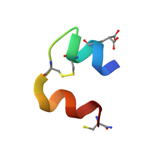
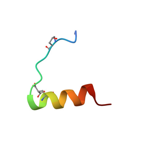
Structure of Conus Geographus insulin G1

  • Classification: HORMONE
  • Organism(s): Conus geographus
  • Mutation(s): No 

  • Deposited: 2016-05-15 Released: 2016-09-07 
  • Deposition Author(s): Lawrence, M.C., Menting, J., Norton, R.S., Safavi-Hemami, H.
  • Funding Organization(s): National Health and Medical Research Council (NHMRC, Australia), European Commission, National Institutes of Health/National Institute of General Medical Sciences (NIH/NIGMS)

Experimental Data Snapshot

  • Method: X-RAY DIFFRACTION
  • Resolution: 1.95 Å
  • R-Value Free: 
    0.233 (Depositor), 0.230 (DCC) 
  • R-Value Work: 
    0.211 (Depositor), 0.211 (DCC) 
  • R-Value Observed: 
    0.212 (Depositor) 

Starting Model: experimental
View more details

wwPDB Validation 3D Report Full Report

Validation slider image for 5JYQ

This is version 1.7 of the entry. See complete history. 

Literature

A minimized human insulin-receptor-binding motif revealed in a Conus geographus venom insulin.

Menting, J.G., Gajewiak, J., MacRaild, C.A., Chou, D.H., Disotuar, M.M., Smith, N.A., Miller, C., Erchegyi, J., Rivier, J.E., Olivera, B.M., Forbes, B.E., Smith, B.J., Norton, R.S., Safavi-Hemami, H., Lawrence, M.C.

(2016) Nat Struct Mol Biol 23: 916-920

  • DOI: https://doi.org/10.1038/nsmb.3292
  • Primary Citation Related Structures: 
    5JYQ

  • PubMed Abstract: 

    Insulins in the venom of certain fish-hunting cone snails facilitate prey capture by rapidly inducing hypoglycemic shock. One such insulin, Conus geographus G1 (Con-Ins G1), is the smallest known insulin found in nature and lacks the C-terminal segment of the B chain that, in human insulin, mediates engagement of the insulin receptor and assembly of the hormone's hexameric storage form. Removal of this segment (residues B23-B30) in human insulin results in substantial loss of receptor affinity. Here, we found that Con-Ins G1 is monomeric, strongly binds the human insulin receptor and activates receptor signaling. Con-Ins G1 thus is a naturally occurring B-chain-minimized mimetic of human insulin. Our crystal structure of Con-Ins G1 reveals a tertiary structure highly similar to that of human insulin and indicates how Con-Ins G1's lack of an equivalent to the key receptor-engaging residue Phe B24 is mitigated. These findings may facilitate efforts to design ultrarapid-acting therapeutic insulins.


  • Organizational Affiliation: 
    • Walter and Eliza Hall Institute of Medical Research, Parkville, Victoria, Australia.

Macromolecule Content 

  • Total Structure Weight: 5.26 kDa 
  • Atom Count: 396 
  • Modeled Residue Count: 43 
  • Deposited Residue Count: 43 
  • Unique protein chains: 2

Macromolecules

Find similar proteins by:  Sequence   |   3D Structure  
Entity ID: 1
MoleculeChains  Sequence LengthOrganismDetailsImage
Insulin 120Conus geographusMutation(s): 0 
UniProt
Find proteins for A0A0B5AC95 (Conus geographus)
Explore A0A0B5AC95 
Go to UniProtKB:  A0A0B5AC95
Entity Groups
Sequence Clusters30% Identity50% Identity70% Identity90% Identity95% Identity100% Identity
UniProt GroupA0A0B5AC95
Sequence Annotations
Expand
Reference Sequence
Find similar proteins by:  Sequence   |   3D Structure  
Entity ID: 2
MoleculeChains  Sequence LengthOrganismDetailsImage
Insulin 1b23Conus geographusMutation(s): 0 
UniProt
Find proteins for A0A0B5A8Q2 (Conus geographus)
Explore A0A0B5A8Q2 
Go to UniProtKB:  A0A0B5A8Q2
Entity Groups
Sequence Clusters30% Identity50% Identity70% Identity90% Identity95% Identity100% Identity
UniProt GroupA0A0B5A8Q2
Sequence Annotations
Expand
Reference Sequence

Small Molecules

Ligands 1 Unique
IDChains Name / Formula / InChI Key2D Diagram3D Interactions
SO4

Query on SO4


Download:Ideal Coordinates CCD File
C [auth A]SULFATE ION
O4 S
QAOWNCQODCNURD-UHFFFAOYSA-L
Modified Residues  3 Unique
IDChains TypeFormula2D DiagramParent
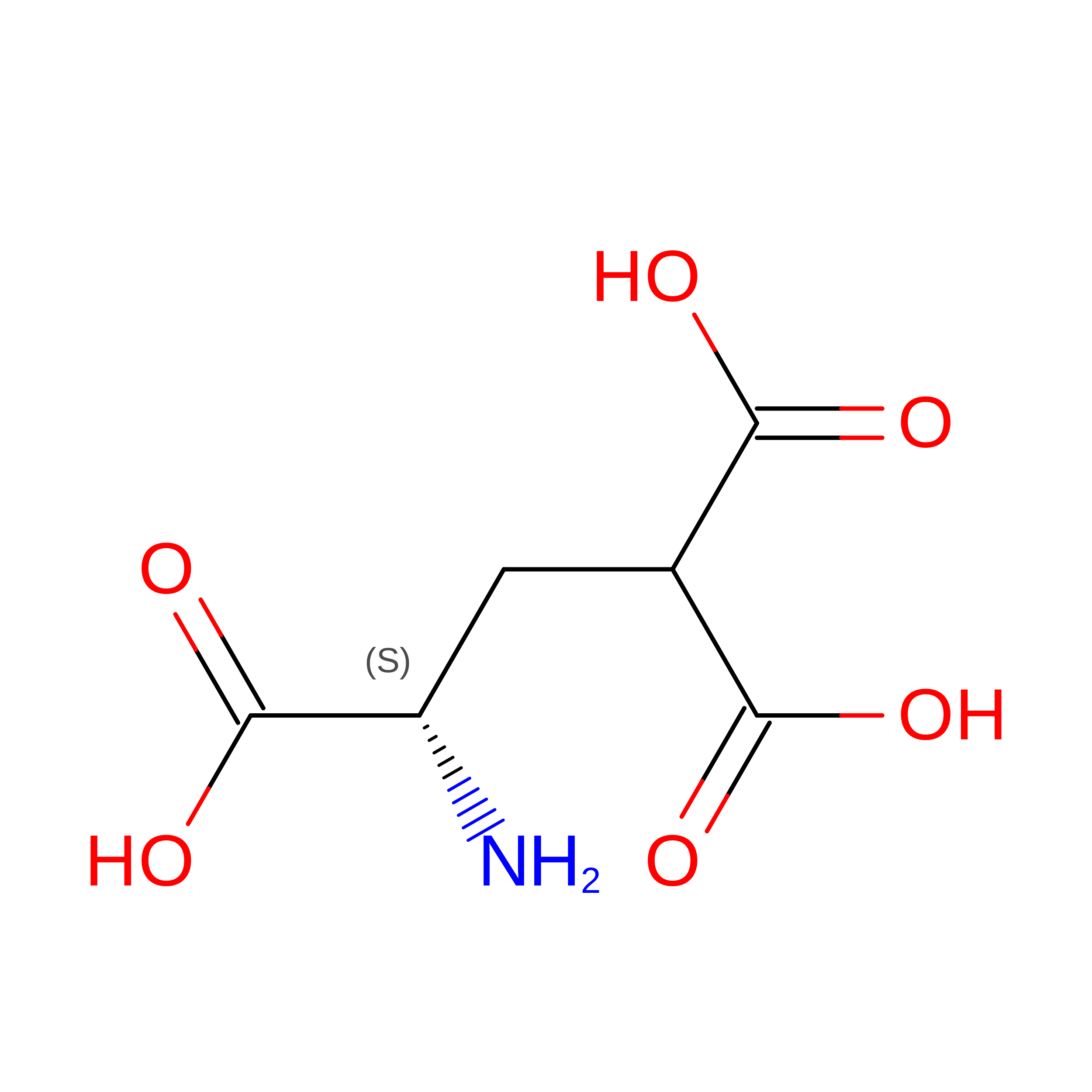
CGU
Query on CGU
A
L-PEPTIDE LINKINGC6 H9 N O6GLU
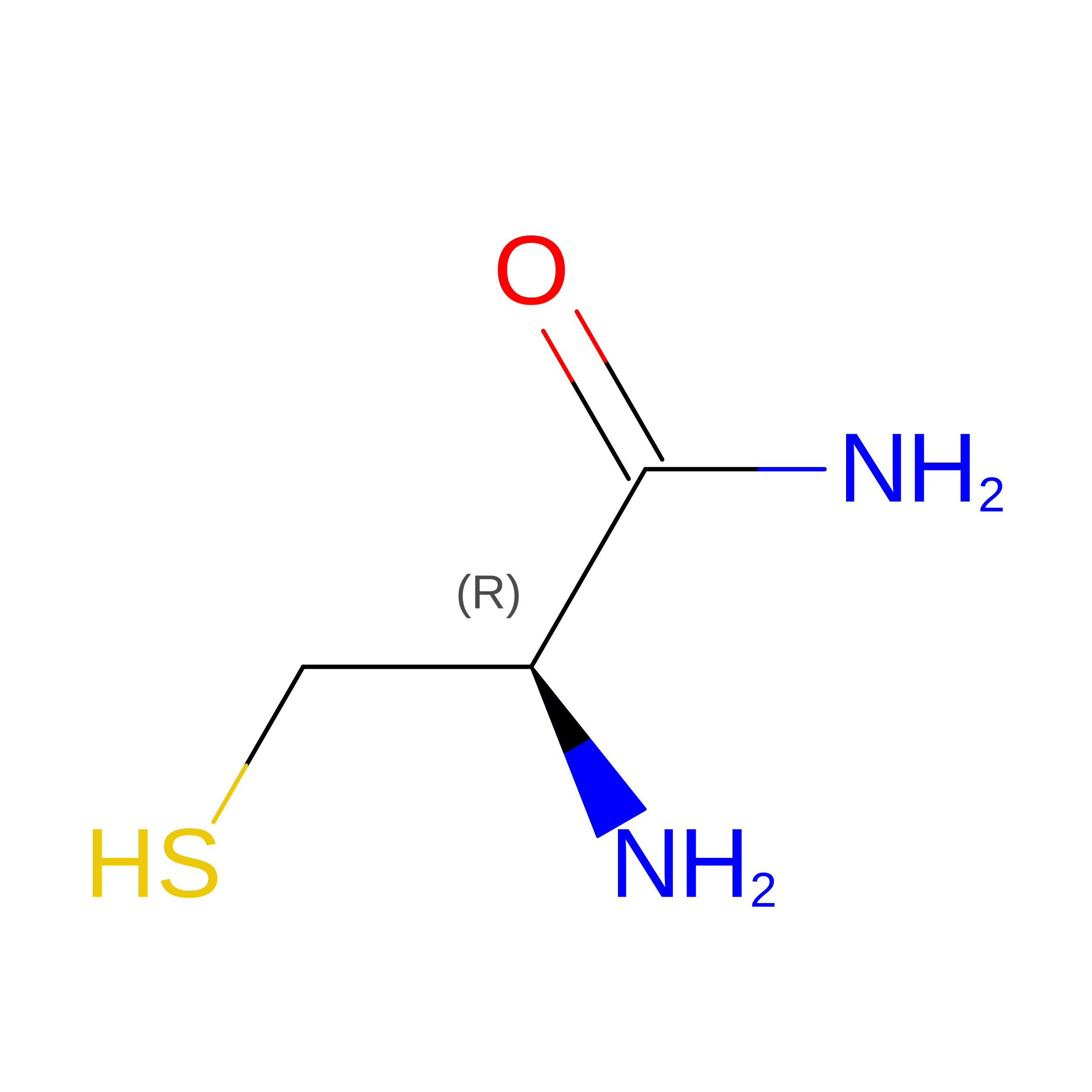
CY3
Query on CY3
A
L-PEPTIDE LINKINGC3 H8 N2 O SCYS
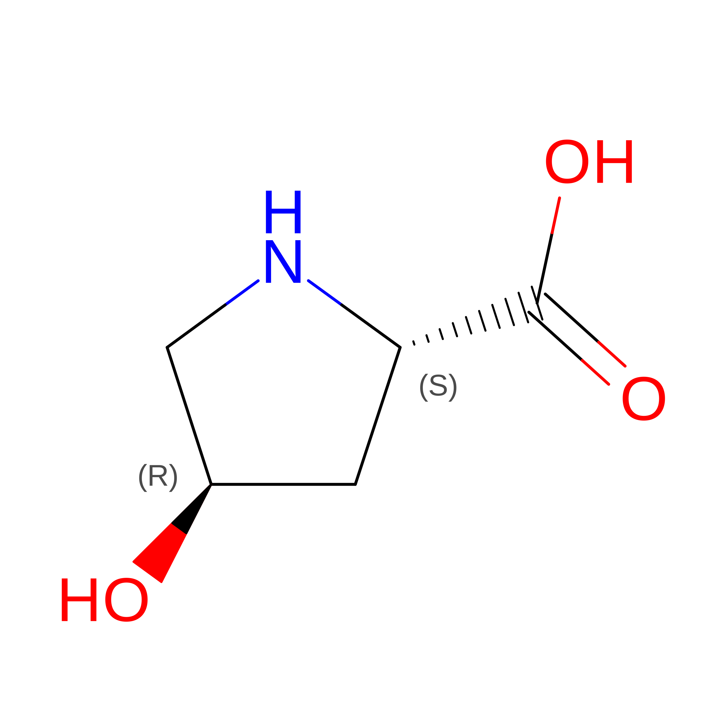
HYP
Query on HYP
B
L-PEPTIDE LINKINGC5 H9 N O3PRO

Experimental Data & Validation

Experimental Data

  • Method: X-RAY DIFFRACTION
  • Resolution: 1.95 Å
  • R-Value Free:  0.233 (Depositor), 0.230 (DCC) 
  • R-Value Work:  0.211 (Depositor), 0.211 (DCC) 
  • R-Value Observed: 0.212 (Depositor) 
Space Group: P 4 3 2
Unit Cell:
Length ( Å )Angle ( ˚ )
a = 74.91α = 90
b = 74.91β = 90
c = 74.91γ = 90
Software Package:
Software NamePurpose
PHENIXrefinement
XDSdata reduction
XDSdata scaling
PHASERphasing

Structure Validation

View Full Validation Report


Entry History 

& Funding Information

Deposition Data


Funding OrganizationLocationGrant Number
National Health and Medical Research Council (NHMRC, Australia)AustraliaAPP1058233
European CommissionCONBIOS 330486
National Institutes of Health/National Institute of General Medical Sciences (NIH/NIGMS)United StatesGM 48677

Revision History  (Full details and data files)

  • Version 1.0: 2016-09-07
    Type: Initial release
  • Version 1.1: 2016-09-14
    Changes: Database references
  • Version 1.2: 2016-09-28
    Changes: Database references
  • Version 1.3: 2016-10-19
    Changes: Database references
  • Version 1.4: 2017-09-20
    Changes: Author supporting evidence, Data collection, Derived calculations
  • Version 1.5: 2019-12-25
    Changes: Author supporting evidence, Derived calculations
  • Version 1.6: 2023-09-27
    Changes: Data collection, Database references, Refinement description
  • Version 1.7: 2023-11-15
    Changes: Data collection