5JIF

Crystal structure of mouse hepatitis virus strain DVIM Hemagglutinin-Esterase


Experimental Data Snapshot

  • Method: X-RAY DIFFRACTION
  • Resolution: 2.00 Å
  • R-Value Free: 0.226 
  • R-Value Work: 0.205 
  • R-Value Observed: 0.206 

wwPDB Validation   3D Report Full Report


Ligand Structure Quality Assessment 


This is version 2.0 of the entry. See complete history


Literature

Coronavirus receptor switch explained from the stereochemistry of protein-carbohydrate interactions and a single mutation.

Bakkers, M.J.Zeng, Q.Feitsma, L.J.Hulswit, R.J.Li, Z.Westerbeke, A.van Kuppeveld, F.J.Boons, G.J.Langereis, M.A.Huizinga, E.G.de Groot, R.J.

(2016) Proc Natl Acad Sci U S A 113: E3111-E3119

  • DOI: https://doi.org/10.1073/pnas.1519881113
  • Primary Citation of Related Structures:  
    5JIF, 5JIL

  • PubMed Abstract: 

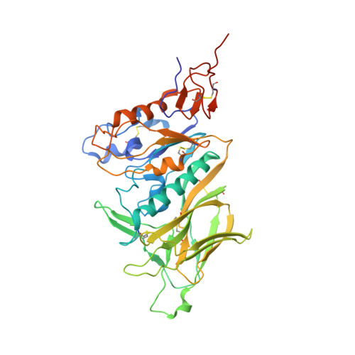
    Hemagglutinin-esterases (HEs) are bimodular envelope proteins of orthomyxoviruses, toroviruses, and coronaviruses with a carbohydrate-binding "lectin" domain appended to a receptor-destroying sialate-O-acetylesterase ("esterase"). In concert, these domains facilitate dynamic virion attachment to cell-surface sialoglycans. Most HEs (type I) target 9-O-acetylated sialic acids (9-O-Ac-Sias), but one group of coronaviruses switched to using 4-O-Ac-Sias instead (type II). This specificity shift required quasisynchronous adaptations in the Sia-binding sites of both lectin and esterase domains. Previously, a partially disordered crystal structure of a type II HE revealed how the shift in lectin ligand specificity was achieved. How the switch in esterase substrate specificity was realized remained unresolved, however. Here, we present a complete structure of a type II HE with a receptor analog in the catalytic site and identify the mutations underlying the 9-O- to 4-O-Ac-Sia substrate switch. We show that (i) common principles pertaining to the stereochemistry of protein-carbohydrate interactions were at the core of the transition in lectin ligand and esterase substrate specificity; (ii) in consequence, the switch in O-Ac-Sia specificity could be readily accomplished via convergent intramolecular coevolution with only modest architectural changes in lectin and esterase domains; and (iii) a single, inconspicuous Ala-to-Ser substitution in the catalytic site was key to the emergence of the type II HEs. Our findings provide fundamental insights into how proteins "see" sugars and how this affects protein and virus evolution.


  • Organizational Affiliation

    Virology Division, Department of Infectious Diseases and Immunology, Faculty of Veterinary Medicine, Utrecht University, 3584 CH Utrecht, The Netherlands;


Macromolecules
Find similar proteins by:  (by identity cutoff)  |  3D Structure
Entity ID: 1
MoleculeChains Sequence LengthOrganismDetailsImage
Hemagglutinin-esterase
A, B
381Murine coronavirus strain DVIMMutation(s): 0 
Gene Names: HE2b
EC: 3.1.1.53
UniProt
Find proteins for O92367 (Murine coronavirus (strain DVIM))
Explore O92367 
Go to UniProtKB:  O92367
Entity Groups  
Sequence Clusters30% Identity50% Identity70% Identity90% Identity95% Identity100% Identity
UniProt GroupO92367
Sequence Annotations
Expand
  • Reference Sequence
Oligosaccharides

Help

Entity ID: 2
MoleculeChains Length2D Diagram Glycosylation3D Interactions
2-acetamido-2-deoxy-beta-D-glucopyranose-(1-4)-2-acetamido-2-deoxy-beta-D-glucopyranose
C, D, E, H, I
C, D, E, H, I, J, K, L
2N-Glycosylation
Glycosylation Resources
GlyTouCan:  G42666HT
GlyCosmos:  G42666HT
GlyGen:  G42666HT
Entity ID: 3
MoleculeChains Length2D Diagram Glycosylation3D Interactions
alpha-D-mannopyranose-(1-2)-alpha-D-mannopyranose-(1-6)-[alpha-D-mannopyranose-(1-3)]alpha-D-mannopyranose-(1-6)-[alpha-D-mannopyranose-(1-3)]beta-D-mannopyranose-(1-4)-2-acetamido-2-deoxy-beta-D-glucopyranose-(1-4)-2-acetamido-2-deoxy-beta-D-glucopyranose
F
8N-Glycosylation
Glycosylation Resources
GlyTouCan:  G61846BY
GlyCosmos:  G61846BY
GlyGen:  G61846BY
Entity ID: 4
MoleculeChains Length2D Diagram Glycosylation3D Interactions
alpha-D-mannopyranose-(1-3)-[alpha-D-mannopyranose-(1-6)]beta-D-mannopyranose-(1-4)-2-acetamido-2-deoxy-beta-D-glucopyranose-(1-4)-2-acetamido-2-deoxy-beta-D-glucopyranose
G
5N-Glycosylation
Glycosylation Resources
GlyTouCan:  G22768VO
GlyCosmos:  G22768VO
GlyGen:  G22768VO
Small Molecules
Ligands 3 Unique
IDChains Name / Formula / InChI Key2D Diagram3D Interactions
NAG
Query on NAG

Download Ideal Coordinates CCD File 
O [auth A],
P [auth A],
Q [auth A],
T [auth B],
U [auth B]
2-acetamido-2-deoxy-beta-D-glucopyranose
C8 H15 N O6
OVRNDRQMDRJTHS-FMDGEEDCSA-N
GOL
Query on GOL

Download Ideal Coordinates CCD File 
N [auth A],
R [auth A]
GLYCEROL
C3 H8 O3
PEDCQBHIVMGVHV-UHFFFAOYSA-N
NA
Query on NA

Download Ideal Coordinates CCD File 
M [auth A],
S [auth B]
SODIUM ION
Na
FKNQFGJONOIPTF-UHFFFAOYSA-N
Experimental Data & Validation

Experimental Data

  • Method: X-RAY DIFFRACTION
  • Resolution: 2.00 Å
  • R-Value Free: 0.226 
  • R-Value Work: 0.205 
  • R-Value Observed: 0.206 
  • Space Group: P 21 21 21
Unit Cell:
Length ( Å )Angle ( ˚ )
a = 88.523α = 90
b = 88.819β = 90
c = 122.163γ = 90
Software Package:
Software NamePurpose
REFMACrefinement
EVAL15data reduction
EVAL15data scaling
PHASERphasing

Structure Validation

View Full Validation Report


Ligand Structure Quality Assessment 


Entry History & Funding Information

Deposition Data


Funding OrganizationLocationGrant Number
Netherlands Organization for Scientific ResearchNetherlandsECHO 711.011.006

Revision History  (Full details and data files)

  • Version 1.0: 2016-05-11
    Type: Initial release
  • Version 1.1: 2016-06-01
    Changes: Database references
  • Version 1.2: 2016-06-08
    Changes: Database references
  • Version 1.3: 2017-09-13
    Changes: Author supporting evidence
  • Version 1.4: 2019-04-03
    Changes: Data collection, Source and taxonomy
  • Version 2.0: 2020-07-29
    Type: Remediation
    Reason: Carbohydrate remediation
    Changes: Atomic model, Data collection, Derived calculations, Structure summary