5HF3

Crystal structure of C-terminal modified Tau peptide-hybrid 201D with 14-3-3sigma


Experimental Data Snapshot

  • Method: X-RAY DIFFRACTION
  • Resolution: 1.80 Å
  • R-Value Free: 0.205 
  • R-Value Work: 0.166 
  • R-Value Observed: 0.169 

wwPDB Validation   3D Report Full Report


This is version 1.2 of the entry. See complete history


Literature

Stabilizer-Guided Inhibition of Protein-Protein Interactions.

Milroy, L.G.Bartel, M.Henen, M.A.Leysen, S.Adriaans, J.M.Brunsveld, L.Landrieu, I.Ottmann, C.

(2015) Angew Chem Int Ed Engl 54: 15720-15724

  • DOI: https://doi.org/10.1002/anie.201507976
  • Primary Citation of Related Structures:  
    4Y32, 4Y3B, 4Y5I, 5HF3

  • PubMed Abstract: 

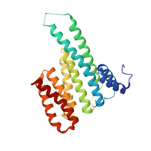
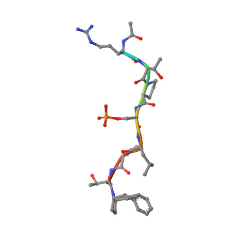
    The discovery of novel protein-protein interaction (PPI) modulators represents one of the great molecular challenges of the modern era. PPIs can be modulated by either inhibitor or stabilizer compounds, which target different though proximal regions of the protein interface. In principle, protein-stabilizer complexes can guide the design of PPI inhibitors (and vice versa). In the present work, we combine X-ray crystallographic data from both stabilizer and inhibitor co-crystal complexes of the adapter protein 14-3-3 to characterize, down to the atomic scale, inhibitors of the 14-3-3/Tau PPI, a potential drug target to treat Alzheimer's disease. The most potent compound notably inhibited the binding of phosphorylated full-length Tau to 14-3-3 according to NMR spectroscopy studies. Our work sets a precedent for the rational design of PPI inhibitors guided by PPI stabilizer-protein complexes while potentially enabling access to new synthetically tractable stabilizers of 14-3-3 and other PPIs.


  • Organizational Affiliation

    Laboratory of Chemical Biology and Institute of Complex Molecular Systems (ICMS), Department of Biomedical Engineering, Technische Universiteit Eindhoven, Den Dolech 2, 5612 AZ Eindhoven (The Netherlands). l.milroy@tue.nl.


Macromolecules
Find similar proteins by:  (by identity cutoff)  |  3D Structure
Entity ID: 1
MoleculeChains Sequence LengthOrganismDetailsImage
14-3-3 protein sigma236Homo sapiensMutation(s): 0 
Gene Names: SFNHME1
UniProt & NIH Common Fund Data Resources
Find proteins for P31947 (Homo sapiens)
Explore P31947 
Go to UniProtKB:  P31947
PHAROS:  P31947
GTEx:  ENSG00000175793 
Entity Groups  
Sequence Clusters30% Identity50% Identity70% Identity90% Identity95% Identity100% Identity
UniProt GroupP31947
Sequence Annotations
Expand
  • Reference Sequence

Find similar proteins by:  Sequence   |   3D Structure  

Entity ID: 2
MoleculeChains Sequence LengthOrganismDetailsImage
modified Tau peptide9synthetic constructMutation(s): 0 
UniProt & NIH Common Fund Data Resources
Find proteins for P10636 (Homo sapiens)
Explore P10636 
Go to UniProtKB:  P10636
PHAROS:  P10636
GTEx:  ENSG00000186868 
Entity Groups  
UniProt GroupP10636
Sequence Annotations
Expand
  • Reference Sequence
Small Molecules
Modified Residues  2 Unique
IDChains TypeFormula2D DiagramParent
CSO
Query on CSO
A
L-PEPTIDE LINKINGC3 H7 N O3 SCYS
SEP
Query on SEP
B
L-PEPTIDE LINKINGC3 H8 N O6 PSER
Experimental Data & Validation

Experimental Data

  • Method: X-RAY DIFFRACTION
  • Resolution: 1.80 Å
  • R-Value Free: 0.205 
  • R-Value Work: 0.166 
  • R-Value Observed: 0.169 
  • Space Group: C 2 2 21
Unit Cell:
Length ( Å )Angle ( ˚ )
a = 82.313α = 90
b = 112.467β = 90
c = 62.43γ = 90
Software Package:
Software NamePurpose
XDSdata reduction
PHASERphasing
PHENIXrefinement
PDB_EXTRACTdata extraction

Structure Validation

View Full Validation Report


Entry History 

Deposition Data

Revision History  (Full details and data files)

  • Version 1.0: 2016-01-27
    Type: Initial release
  • Version 1.1: 2017-09-13
    Changes: Advisory, Data collection
  • Version 1.2: 2024-01-10
    Changes: Advisory, Data collection, Database references, Derived calculations, Refinement description