5HBU

Structure of the E. coli nucleoid occlusion protein SlmA bound to DNA and the C-terminal tail of the cytoskeletal cell division protein FtsZ


Experimental Data Snapshot

  • Method: X-RAY DIFFRACTION
  • Resolution: 2.60 Å
  • R-Value Free: 0.270 
  • R-Value Work: 0.226 
  • R-Value Observed: 0.228 

wwPDB Validation   3D Report Full Report


This is version 1.3 of the entry. See complete history


Literature

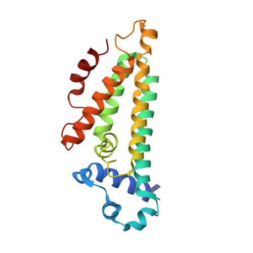
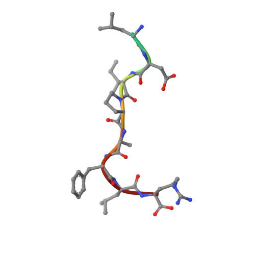
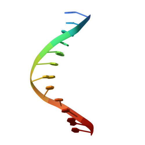
Structures of the nucleoid occlusion protein SlmA bound to DNA and the C-terminal domain of the cytoskeletal protein FtsZ.

Schumacher, M.A.Zeng, W.

(2016) Proc Natl Acad Sci U S A 113: 4988-4993

  • DOI: https://doi.org/10.1073/pnas.1602327113
  • Primary Citation of Related Structures:  
    5HAW, 5HBU, 5HSZ, 5K58

  • PubMed Abstract: 

    Cell division in most prokaryotes is mediated by FtsZ, which polymerizes to create the cytokinetic Z ring. Multiple FtsZ-binding proteins regulate FtsZ polymerization to ensure the proper spatiotemporal formation of the Z ring at the division site. The DNA-binding protein SlmA binds to FtsZ and prevents Z-ring formation through the nucleoid in a process called "nucleoid occlusion" (NO). As do most FtsZ-accessory proteins, SlmA interacts with the conserved C-terminal domain (CTD) that is connected to the FtsZ core by a long, flexible linker. However, SlmA is distinct from other regulatory factors in that it must be DNA-bound to interact with the FtsZ CTD. Few structures of FtsZ regulator-CTD complexes are available, but all reveal the CTD bound as a helix. To deduce the molecular basis for the unique SlmA-DNA-FtsZ CTD regulatory interaction and provide insight into FtsZ-regulator protein complex formation, we determined structures of Escherichia coli, Vibrio cholera, and Klebsiella pneumonia SlmA-DNA-FtsZ CTD ternary complexes. Strikingly, the FtsZ CTD does not interact with SlmA as a helix but binds as an extended conformation in a narrow, surface-exposed pocket formed only in the DNA-bound state of SlmA and located at the junction between the DNA-binding and C-terminal dimer domains. Binding studies are consistent with the structure and underscore key interactions in complex formation. Combined, these data reveal the molecular basis for the SlmA-DNA-FtsZ interaction with implications for SlmA's NO function and underscore the ability of the FtsZ CTD to adopt a wide range of conformations, explaining its ability to bind diverse regulatory proteins.


  • Organizational Affiliation

    Department of Biochemistry, Duke University School of Medicine, Durham, NC 27710 maria.schumacher@duke.edu.


Macromolecules

Find similar proteins by:  (by identity cutoff)  |  3D Structure
Entity ID: 1
MoleculeChains Sequence LengthOrganismDetailsImage
Nucleoid occlusion factor SlmA
A, B, C, D, E
A, B, C, D, E, F, G, H
196Escherichia coli K-12Mutation(s): 0 
Gene Names: slmAttkyicBb3641JW5641
UniProt
Find proteins for P0C093 (Escherichia coli (strain K12))
Explore P0C093 
Go to UniProtKB:  P0C093
Entity Groups  
Sequence Clusters30% Identity50% Identity70% Identity90% Identity95% Identity100% Identity
UniProt GroupP0C093
Sequence Annotations
Expand
  • Reference Sequence

Find similar proteins by:  Sequence   |   3D Structure  

Entity ID: 3
MoleculeChains Sequence LengthOrganismDetailsImage
FtsZ CTT peptideM [auth K]10synthetic constructMutation(s): 0 
UniProt
Find proteins for P0A9A6 (Escherichia coli (strain K12))
Explore P0A9A6 
Go to UniProtKB:  P0A9A6
Entity Groups  
Sequence Clusters30% Identity50% Identity70% Identity90% Identity95% Identity100% Identity
UniProt GroupP0A9A6
Sequence Annotations
Expand
  • Reference Sequence

Find similar nucleic acids by:  Sequence   |   3D Structure  

Entity ID: 2
MoleculeChains LengthOrganismImage
DNA (5'-D(*GP*TP*GP*AP*GP*TP*AP*CP*TP*CP*AP*C)-3')I [auth W],
J [auth Z],
K [auth R],
L [auth T]
12synthetic construct
Sequence Annotations
Expand
  • Reference Sequence
Experimental Data & Validation

Experimental Data

  • Method: X-RAY DIFFRACTION
  • Resolution: 2.60 Å
  • R-Value Free: 0.270 
  • R-Value Work: 0.226 
  • R-Value Observed: 0.228 
  • Space Group: P 21 21 21
Unit Cell:
Length ( Å )Angle ( ˚ )
a = 70.114α = 90
b = 158.893β = 90
c = 200.201γ = 90
Software Package:
Software NamePurpose
PHENIXrefinement
MOSFLMdata reduction
SCALAdata scaling
MOLREPphasing
PDB_EXTRACTdata extraction

Structure Validation

View Full Validation Report


Entry History 

Deposition Data

Revision History  (Full details and data files)

  • Version 1.0: 2016-04-13
    Type: Initial release
  • Version 1.1: 2016-05-04
    Changes: Database references
  • Version 1.2: 2016-05-18
    Changes: Database references
  • Version 1.3: 2023-09-27
    Changes: Data collection, Database references, Derived calculations, Refinement description