5FLO | pdb_00005flo

Native state mass spectrometry, surface plasmon resonance and X-ray crystallography correlate strongly as a fragment screening combination


Experimental Data Snapshot

  • Method: X-RAY DIFFRACTION
  • Resolution: 1.66 Å
  • R-Value Free: 
    0.215 (Depositor), 0.226 (DCC) 
  • R-Value Work: 
    0.158 (Depositor), 0.172 (DCC) 
  • R-Value Observed: 
    0.161 (Depositor) 

Starting Model: experimental
View more details

wwPDB Validation 3D Report Full Report

Validation slider image for 5FLO

Ligand Structure Quality Assessment 


This is version 1.3 of the entry. See complete history

Literature

Native State Mass Spectrometry, Surface Plasmon Resonance and X-Ray Crystallography Correlate Strongly as a Fragment Screening Combination.

Woods, L.Dolezal, O.Ren, B.Ryan, J.H.Peat, T.S.Poulsen, S.

(2016) J Med Chem 59: 2192

  • DOI: https://doi.org/10.1021/acs.jmedchem.5b01940
  • Primary Citation Related Structures: 
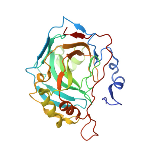
    5EH5, 5EH7, 5EH8, 5EHV, 5EHW, 5FLO, 5FLP, 5FLQ, 5FLR, 5FLS, 5FLT, 5FNG, 5FNH, 5FNI, 5FNJ, 5FNK, 5FNL, 5FNM

  • PubMed Abstract: 

    Fragment-based drug discovery (FBDD) is contingent on the development of analytical methods to identify weak protein-fragment noncovalent interactions. Herein we have combined an underutilized fragment screening method, native state mass spectrometry, together with two proven and popular fragment screening methods, surface plasmon resonance and X-ray crystallography, in a fragment screening campaign against human carbonic anhydrase II (CA II). In an initial fragment screen against a 720-member fragment library (the "CSIRO Fragment Library") seven CA II binding fragments, including a selection of nonclassical CA II binding chemotypes, were identified. A further 70 compounds that comprised the initial hit chemotypes were subsequently sourced from the full CSIRO compound collection and screened. The fragment results were extremely well correlated across the three methods. Our findings demonstrate that there is a tremendous opportunity to apply native state mass spectrometry as a complementary fragment screening method to accelerate drug discovery.


  • Organizational Affiliation
    • Griffith University , Eskitis Institute for Drug Discovery, Brisbane, Queensland Australia.

Macromolecule Content 

  • Total Structure Weight: 29.57 kDa 
  • Atom Count: 2,405 
  • Modeled Residue Count: 258 
  • Deposited Residue Count: 260 
  • Unique protein chains: 1

Macromolecules

Find similar proteins by:|  3D Structure
Entity ID: 1
MoleculeChains  Sequence LengthOrganismDetailsImage
CARBONIC ANHYDRASE 2260Homo sapiensMutation(s): 0 
EC: 4.2.1.1 (PDB Primary Data), 4.2.1.69 (UniProt)
UniProt & NIH Common Fund Data Resources
Find proteins for P00918 (Homo sapiens)
Explore P00918 
Go to UniProtKB:  P00918
PHAROS:  P00918
GTEx:  ENSG00000104267 
Entity Groups
Sequence Clusters30% Identity50% Identity70% Identity90% Identity95% Identity100% Identity
UniProt GroupP00918
Sequence Annotations
Expand
Reference Sequence

Small Molecules

Ligands 2 Unique
IDChains Name / Formula / InChI Key2D Diagram3D Interactions
J4K

Query on J4K


Download:Ideal Coordinates CCD File
C [auth A]5-[(4-chloranylphenoxy)methyl]-1H-1,2,3,4-tetrazole
C8 H7 Cl N4 O
LAEFYQOLGWVFTE-UHFFFAOYSA-N
ZN

Query on ZN


Download:Ideal Coordinates CCD File
B [auth A]ZINC ION
Zn
PTFCDOFLOPIGGS-UHFFFAOYSA-N

Experimental Data & Validation

Experimental Data

  • Method: X-RAY DIFFRACTION
  • Resolution: 1.66 Å
  • R-Value Free:  0.215 (Depositor), 0.226 (DCC) 
  • R-Value Work:  0.158 (Depositor), 0.172 (DCC) 
  • R-Value Observed: 0.161 (Depositor) 
Space Group: P 1 21 1
Unit Cell:
Length ( Å )Angle ( ˚ )
a = 42.753α = 90
b = 41.493β = 104.6
c = 71.66γ = 90
Software Package:
Software NamePurpose
REFMACrefinement
XDSdata reduction
Aimlessdata scaling
PHASERphasing

Structure Validation

View Full Validation Report


Ligand Structure Quality Assessment 


Entry History 

Deposition Data

Revision History  (Full details and data files)

  • Version 1.0: 2016-03-02
    Type: Initial release
  • Version 1.1: 2016-03-23
    Changes: Database references
  • Version 1.2: 2019-05-08
    Changes: Data collection, Experimental preparation, Other
  • Version 1.3: 2024-01-10
    Changes: Data collection, Database references, Derived calculations, Other, Refinement description